
Page 126 - Basic Principles of Textile Coloration
P. 126
STRUCTURE OF WOOL FIBRES 115
pigments. It does not run the entire length of the fibre and there may be hollow
spaces.
A model for dyeing based on transfer of the dye from the aqueous solution to
the fibre surface, adsorption on the surface and diffusion into the fibre, seems
simplistic on considering the complex morphology of the wool fibre. The wool
cortex is far from being homogeneous. Different parts of a wool fibre have different
degrees of dye absorption due to variations in permeability and chemical
composition. Studies on the rate of diffusion of dyes into wool fibres indicate a
surface barrier opposing diffusion. This is particularly evident in the early stages of
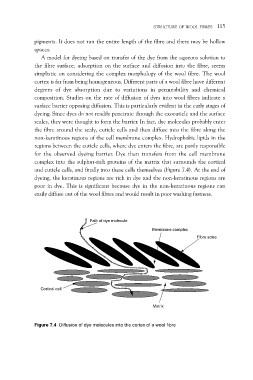
dyeing. Since dyes do not readily penetrate through the exocuticle and the surface
scales, they were thought to form the barrier. In fact, dye molecules probably enter
the fibre around the scaly, cuticle cells and then diffuse into the fibre along the
non-keratinous regions of the cell membrane complex. Hydrophobic lipids in the
regions between the cuticle cells, where dye enters the fibre, are partly responsible
for the observed dyeing barrier. Dye then transfers from the cell membrane
complex into the sulphur-rich proteins of the matrix that surrounds the cortical
and cuticle cells, and finally into these cells themselves (Figure 7.4). At the end of
dyeing, the keratinous regions are rich in dye and the non-keratinous regions are
poor in dye. This is significant because dye in the non-keratinous regions can
easily diffuse out of the wool fibres and would result in poor washing fastness.
Path of dye molecule
Membrane complex
Fibre scale
Cortical cell
Matrix
Figure 7.4 Diffusion of dye molecules into the cortex of a wool fibre