
Page 1157 - Hall et al (2015) Principles of Critical Care-McGraw-Hill
P. 1157
796 PART 6: Neurologic Disorders
The degree of tissue displacement, and therefore a prediction of
TABLE 86-4 Herniation Syndromes (Continued)
compressed structures and expected symptoms, can be approximated
Herniation Mechanism/Imaging Bedside Examination/ by measuring the horizontal shift of the calcified pineal gland on a
Syndromes Findings Comments noncontrast head CT. In a classic study describing this important rela-
Transdural/ • Increased ICP forces brain through • Sometimes called “brain tionship, horizontal shift of the pineal gland from its midline position
transcranial dura ± subgaleal extension fungus” (eg, Fig. 86-12B) by 0 to 3 mm correlated with wakefulness; 3 to 5 mm
28
herniation • Brain and vessels herniated through • Used as therapeutic option with drowsiness; 6 to 8 mm with stupor; and >8 mm with coma. In
dural ± skull defect to decompress swollen comparison, rostrocaudal displacement leads to herniation of brain
brain (“hemicraniectomy”) structures through the foramen magnum with resultant pressure on the
• Use changes in turgor of dorsal brainstem and obstruction of CSF outflow providing two mecha-
“pseudofontanelle” for nisms for depressed consciousness: direct injury to anatomic structures
serial bedside examination responsible for arousal and elevated ICP via hydrocephalus. Midline
• Too small craniectomy shift is often measured as the distance from the falx (midline) to the sep-
can lead to bleeding and tum pellucidum and a ratio can be calculated (Figs. 86-12 and 86-13). 29
brain injury at the bony Notably, the extent of horizontal shift seen on imaging is not always
edges the primary mechanism of a patient’s reduced level of consciousness.
Other differential etiologies should always be considered. Further, the
Descending Type
clinical sequelae of tissue displacement and herniation vary greatly
Central or • Both temporal lobes herniate into • Impaired consciousness among patients due to underlying factors that alter reserve and com-
bilateral tentorial opening with associated pliance. As an example, an atrophic brain with significantly increased
downward • Optic chiasm/diencephalon oculopupillomotor subarachnoid spaces possesses a greater ability to compensate for tissue
transtentorial compressed against skull base changes displacement by decreasing subarachnoid space to maintain a constant
herniation • Midbrain displaced inferiorly: • Progressive loss of all pressure. As a result, an individual’s reserve factors contributing to
anterior inferior third ventricle brainstem function compliance must be taken into consideration when assessing the rela-
displaced posteriorly behind dorsum; leading to brain death tionship between radiographic and clinical presentations in the setting
sella angle between midbrain • Bilateral flexor or of intracranial mass lesions and tissue displacement. Some sequelae of
and pons becomes more acute extensor posturing from herniation are summarized in Table 86-4.
(brainstem budding) progressive brainstem
• Complications: injury
Penetrating basal arteries
occlusion brainstem infarcts
and hemorrhages (Duret)
Hydrocephalus
Tonsillar • Tonsils pushed inferiorly into • Common with posterior
foramen magnum as displacement fossa masses
>5 mm and tonsil folia become • Disturbance of conjugate
vertically oriented gaze, quadriparesis,
• Cisterna magna obliterates autonomic symptoms
• Complications: (changes in blood pressure
Fourth ventricle outlet obstruction and heart rate)
produces hydrocephalus • Miosis and ataxic
Compression of medulla produces breathing
changes in respiration • Severe and progressive
and cardiovascular homeostasis headache with associated
nausea and vomiting
• Sometimes early
sign of obstructive
hydrocephalus
Ascending Type
Transtentorial • Infratentorial mass lesion pushes • Less common than
cerebellum and upper brainstem descending herniation
upward into the tentorial opening • Can be caused by a slowly
• Subsequent narrowing of the bilateral growing cerebellar or
ambient cisterns as the cerebellar tissue brainstem process, such
extends into the ambient cisterns as diffusely infiltrating
• Quadrigeminal cistern closed, tectum astrocytoma
flattening • Nausea and vomiting are
• Aqueduct obstruction leading to commonly seen followed
obstructive hydrocephalus by obtundation and coma
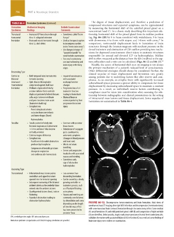
depending on the length FIGURE 86-12. Decompressive hemicraniectomy and brain herniation. Axial views of
of time the mass effect unenhanced head CT imaging after right MCA infarction and decompression (hemicraniectomy)
has been present in the with some transdural brain (release) herniation through the craniectomy defect. Some residual
posterior fossa (A) uncal herniation (A) and mild persistent pineal shift (B) and compression of right ventricle
(D) are identified. Unfortunately, despite early decompression with small hemicraniectomy site,
CPA, cerebellopontine angle; ICP, intracranial pressure. subfalcine herniation with associated bilateral ACA infarction (C) occurred and some findings of
Herniation syndrome categorization and clinical-imaging correlations are provided. brainstem injury were evident on examination.
section06.indd 796 1/23/2015 12:55:54 PM