
Page 1158 - Hall et al (2015) Principles of Critical Care-McGraw-Hill
P. 1158
CHAPTER 86: Intracranial Pressure: Monitoring and Management 797
from a medially displaced uncus, which subsequently evolves to stupor,
coma, and contralateral pupil dilation from further brainstem compres-
sion if the source of the herniation is not corrected. Pupillary changes can
reverse with successful, rapid ICP normalization. Uncal herniation can
result in trapping of the ipsilateral temporal horn of the lateral ventricle
with resultant CSF obstruction, dilation of the temporal horn and sur-
rounding tissue (Fig. 86-12).
Central transtentorial herniation (Fig. 86-11-2) is most common with
global, bihemispheric processes (eg, global ischemia/infarction, menin-
gitis, or fulminant hepatic failure) and it classically occurs as the cerebral
hemispheres and basal ganglia exert downward pressure, causing brain
displacement through the tentorial incisura bilaterally with the pressure
cone into the brainstem. If progressive, it results in severe brainstem
compression and ischemia with hemorrhage. Bilateral PCA compression
can occur with resulting ischemia of the PCA territories as well as the
potential for CSF outflow obstruction with hydrocephalus.
In comparison to supratentorial lesions, a posterior fossa mass
exerts direct pressure on the brainstem from downward displacement
of the cerebellar tonsils (tonsillar herniation) and lower brainstem
(medulla) through the foramen magnum (Fig. 86-11-6). This causes
severe brainstem and upper cervical spinal cord compression, as well as
obstruction of CSF outflow resulting in hydrocephalus. Clinically, these
patients develop symptoms of brainstem dysfunction such as autonomic
disturbance, altered respiratory patterns, pyramidal tract signs, and
cranial nerve palsies as well as depressed consciousness. In addition to
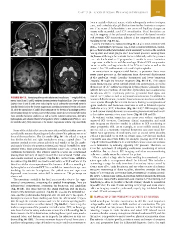
FIGURE 86-13. Herniation pathways with infratentorial mass lesions. T1-weighted MRI of a downward displacement, posterior fossa lesions can also force cerebellar
normal brain (A and C) and T2-weighted abnormal posterior fossa lesion (B and D) are presented. tissue upward through the tentorial incisura, leading to compression of
Sagittal views (A and B) with arrow indicating the typical pathway for downward cerebellar upper cerebellar and brainstem structures as well as bilateral superior
(tonsillar) herniation into the foramen magnum and ascending transtentorial herniation is shown cerebellar artery (SCA) infarctions (Fig. 86-11-5). MR imaging (sagit-
30
(B), while the coronal views (C and D) clearly demonstrate the direction of ascending transtento- tal and coronal views) demonstrates ascending transtentorial and tonsil-
rial herniation through the opening. Acute cerebellar mass lesions (ie, tumors) can readily induce lar herniation secondary to a mass lesion (Fig. 86-13B and D).
these cerebellar herniation syndromes as well as lead to brainstem compression, obstructive As outlined earlier, herniation can occur even without significant
hydrocephalus, and ischemic infarctions from posterior inferior cerebellar artery (PICA) and supe- measured ICP elevation. Continuous clinical examination and serial
rior cerebellar artery (SCA) compression at the foramen magnum and tentorial edge, respectively.
brain imaging are therefore needed in addition to ICP monitoring to
detect progressive shift. As an example, an acute middle cranial fossa
Some of the deficits that occur in association with herniation evolve in process such as a traumatic temporal hematoma can cause uncal her-
a predictable manner depending on the location of the primary vector of niation with symptoms of local injury, such as cranial nerve disorder,
force of the mass lesion. The falx cerebri (Fig. 86-1) is a dural structure without a profound rise in ICP. In certain cases, ICP-directed medical
that divides the left and right hemispheres along a sagittal plane. The treatments can exacerbate BTD; for example, placing an EVD into a
anterior cerebral arteries course inferiorly and parallel to the falx cerebri trapped ventricle contralateral to a hemispheric mass lesion can increase
and supply blood to the anterior, inferior, and medial frontal lobes. With lateral herniation by relieving opposing CSF pressure. Therefore, we
anterior BTD, brain parenchyma herniates under and across the falx stress the importance of integrating continuous monitoring of several
(subfalcine herniation). The anterior cerebral arteries are compressed, modalities, that is, clinical, ICP, imaging, and other neuromonitoring
placing their territory of supply (mostly the inferomedial frontal lobes tools to accurately assess the status of the patient.
and caudate nucleus) in jeopardy (Fig. 86-12). Furthermore, subfalcine When a patient at high risk for brain swelling is encountered, a pro-
herniation (Fig. 86-10C) can lead to obstruction of CSF outflow of the active approach to management should be initiated. This includes a
lateral ventricles via compression of the foramen of Monro, resulting in monitoring strategy for early detection of secondary injury caused by
hydrocephalus and elevated ICP. Since subfalcine herniation is a process edema, mass effect, brain herniation, and any other sources of ischemia.
involving the anterior hemispheres, the patient may not experience Proactive monitoring of these variables, therefore, provides the best
depressed consciousness unless shift is extreme or CSF pathways are means of detecting and correcting them, preemptively avoiding second-
obstructed. ary injury. As mentioned before, monitoring methods include the physical
The tentorium cerebelli is the dural structure that divides the supra- examination, radiographic assessment, and invasive ICP monitoring. ICP
tentorial compartment, containing the cerebral hemispheres, from the monitoring as close as possible to the site of injury should be considered
infratentorial compartment, containing the brainstem and cerebellum especially when the risk of brain swelling is very high and serial exami-
(Fig. 86-1B). The space between the lateral midbrain and the medial nation or imaging cannot be performed properly (eg, intubated, heavily
border of the tentorium cerebelli (Fig. 86-11) is called the tentorial inci- sedated, difficult to transport).
hemispheric mass or brain swelling can force the inferomedial temporal ■ EXAMINATION OF THE PATIENT WITH SUSPECTED ICP ELEVATION
sura, and the posteromedial temporal lobe sits just above this space. A
lobe through the tentorial incisura and into the tentorial opening (called Serial neurological bedside examination is still the most important,
lateral transtentorial or uncal herniation) (Fig. 86-11-1). Commonly, this indispensable, and readily available method of examination. The pri-
leads to compression of the posterior cerebral arteries (PCA) as they origi- mary drawback to this process, however, is that it can be limiting in
nate from the basilar artery and course around the midbrain (Fig. 86-1). detecting changes in brain function and raised ICP. Altered mental
Brain tissue in the PCA distribution, including the occipital lobes, medial status may be due to many etiologies not limited to elevated ICP, and this
temporal lobes, and thalami, are in jeopardy for infarction in this syn- distinction is impossible to make based on physical examination alone.
drome (Fig. 86-12D). The most common feature of uncal herniation is The patient’s risk factor profile, activity at onset, and tempo of symptom
pupillary enlargement, a sign of third nerve and/or midbrain compression onset and progression limit the differential diagnosis, that is, the rapid
section06.indd 797 1/23/2015 12:55:55 PM