
Page 271 - Hall et al (2015) Principles of Critical Care-McGraw-Hill
P. 271
CHAPTER 26: Therapeutic Hypothermia 175
hypothermia was in the setting of malignancy. In 1939, Fay and col- hypothermic protection include modulation of transcription and/or
leagues treated patients with metastatic carcinoma, with the goal of both translation, suppression of reactive oxygen species (ROS) production,
13
pain reduction and retardation of tumor growth. While hypothermia and inhibition of programmed cell death, or apoptosis. The mechanisms
to 32°C for 24 hours did not prove effective for the stated goals, it was by which cooling protects tissues may overlap as well. For example, the
considered well tolerated. 13 effect of hypothermia on cellular metabolism may lead indirectly to
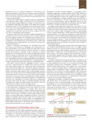
A decade later, Wilfred Bigelow studied the induction of hypothermia modulation of programmed cell death, and cooling may have a direct
in the setting of cardiac surgery, with the goal of cerebral protection. effect on cell death machinery itself (see Fig. 26-1). Most of the data
14
Two other studies using hypothermia as therapy for cardiac arrest were pertaining to mechanism of hypothermic protection comes from studies
also published. Both these early cardiac arrest studies used moderate of IR injury in models of stroke and myocardial infarction.
hypothermia of 30°C to 34°C in patients after resuscitation from cardiac A number of gross physiologic changes have been observed in the
arrest. One of these pioneering papers presented a series of four patients, setting of hypothermia that may contribute to decreased injury. As early
all of whom were cooled and survived arrest. In the other study, as 1954 it was noted that hypothermia induced by ice water immersion
15
12 patients were cooled with a survival rate of 50% compared with 14% could lower cerebral oxygen consumption in dogs by approximately
survival in 7 normothermic control patients. 16 7% per 1°C drop in temperature. Other studies have demonstrated that
29
During the 1960s and 1970s, the field of induced hypothermia lay mild hypothermia in rats improves postischemic cerebral blood flow
relatively dormant for reasons that remain unclear. Some have suggested disturbances. Another marker of general physiologic injury after reper-
30
that more dramatic therapies were developed that overshadowed cool- fusion, brain edema, was also found to be reduced by hypothermia in a
ing as a possible therapy, such as controlled ventilation, monitored ICU rat model of global ischemia. 30,31 Finally, hypothermia has been shown to
management, and cardiopulmonary resuscitation (CPR). Additionally, minimize damage to the blood-brain barrier, which in turn may protect
5
several adverse effects of hypothermia were described, which may have against blood-borne toxic metabolites reaching brain tissues through the
dampened enthusiasm. 17,18 compromised barrier. 30,32
Interest in “resuscitative hypothermia” was rekindled by Peter Safar Intracellular signaling can alter the array of gene transcription activity
and others at the University of Pittsburgh, who demonstrated in a of a cell quickly and dramatically, and this, in turn, can trigger a variety
ventricular fibrillation dog model of cardiac arrest that mild to moder- of injury processes. In a cardiac arrest mouse model, a group of signaling
ate hypothermia could be induced to improve outcomes. 19,20 Trauma pathway genes known as the immediate early genes was activated after
research also provided a motivation for the development of induced resuscitation. A study of liver IR demonstrated a drop in c-jun terminal
33
34
hypothermia. It was understood from military combat experience that kinase activity at 25°C when compared with normothermic controls.
definitive therapy for penetrating trauma was often delayed for practical An extracellular signaling molecule thought to protect against injury,
reasons (eg, transportation and access to surgical care) and that mea- BDNF, was increased in a rat model of cardiac arrest when animals were
sures were needed to preserve exsanguinating soldiers until appropriate cooled to 33°C. 35
care could be delivered. Given the animal data on exsanguination and A number of biochemical changes during IR can be modified by the
21
cooling, it appeared that hypothermia might be a suitable approach. 22 induction of hypothermia. In a gerbil stroke model, animals subjected
Safar went on further to describe “suspended animation,” a process to mild hypothermia were found to have decreased arachidonic acid
that allows “rapid preservation of viability of the organisms in tempo- metabolism compared with normothermic controls. In a rat brain
36
rarily unresuscitable cardiac arrest, which allows time for transport and ischemia model, hypothermia to 32°C reduced nitric oxide produc-
repair during clinical death and is followed by delayed resuscitation, tion, as measured in jugular blood. Whether these attenuations are
37
hopefully to survival without brain damage.” Hypothermia has been a simply markers of hypothermic effects or actually relevant factors in
12
primary component of this concept of stasis. In this paradigm, victims of reperfusion injury remains to be clearly established. Other biochemical
cardiac arrest may be cooled to some target temperature and maintained phenomena seem more likely to be linked directly to damage processes,
at that temperature for a specific period of time. With advanced medical such as the observation that hypothermia slows ATP depletion during
interventions, which may include cardiopulmonary bypass, metabolic IR. ROS production also appears to be attenuated by hypothermic
38
correction, and controlled reperfusion, the patient is stabilized and conditions in a rat cerebral ischemia model. 39
rewarmed, and “reanimation” is initiated. While many methodologies Programmed cell death is a complex yet ubiquitous process by which
have been studied under the rubric of suspended animation, including cells actively chose or are chosen to die. This cellular program can
cardiopulmonary bypass and pharmacologic interventions, these are be activated as part of normal physiology, such as during embryonic
23
used most often as adjuncts to the use of hypothermia. development, or as an abnormal response in a wide variety of disease
Since these initial observations in the 1980s and early 1990s, much of states. 40,41 Much evidence implicates the induction of apoptosis as a
the work pertaining to hypothermia and ischemic disease has focused component of reperfusion injury. 42,43 A recent report showed that the
on focal ischemia and reperfusion, for example, animal stroke and apoptotic pathway enzyme caspase 3 was upregulated in brain tissue
myocardial infarction models. A number of ischemia-reperfusion (IR)
model systems have been developed over the last two decades, including
cellular, isolated organ, and whole-animal models in which arterial Ischemia Hypoxia Reperfusion
24
25
supply to the organ under study is temporarily occluded. 26,27 In this latter
category are included experiences with human IR, for example, during
coronary vascular procedures. More recently, two seminal papers were
28
published describing the use of hypothermia to successfully treat resus-
2,3
citated cardiac arrest patients. With these studies, hypothermia has Reactive Oxygen Inflammatory Mitochondrial Hypothermia
moved from the laboratory to active clinical use. Species (ROS) Cascades Dysfunction
MECHANISMS OF HYPOTHERMIC PROTECTION
Hypotension
The mechanisms by which induced hypothermia protects against cel- Apoptosis
lular and tissue injury are poorly understood. Given the importance of Organ Dysfunction
temperature in a wide range of physiologic processes, it is reasonable
to conclude that multiple mechanisms may be involved in any given FIGURE 26-1. Mechanism of hypothermia may lessen the effects of reperfusion injury,
tissue (reviewed in refs. 5 and 6). Some mechanisms implicated in damage observed after restoration of blood flow to ischemic tissues.
section02.indd 175 1/13/2015 2:05:14 PM