
Page 306 - Hall et al (2015) Principles of Critical Care-McGraw-Hill
P. 306
210 PART 2: General Management of the Patient
■ INDICATIONS AND PATIENT SELECTION contents into sterile cavities. Conscious sedation is preferred, though
Percutaneous drainage is the treatment of choice for abscesses and other in select circumstances, general anesthesia may be necessary. In some
patients, the procedure can be performed with local anesthetic only.
fluid collections such as urinomas and bilomas. Compared with surgical
exploration, percutaneous approaches are less invasive and associated A thorough review of imaging studies will determine the safest access
route. The best route is usually the shortest and straightest pathway.
with decreased mortality. In some instances, percutaneous approaches
1
are less costly. Percutaneous drainage is particularly favored in critically Ideally, the catheter is placed in a convenient location for ongoing care. In
solid organ collections, a small amount of normal parenchyma is traversed
ill patients as they are often not surgical candidates.
When an abscess is suspected in an ICU patient, cross-sectional imag- to aid fixation and mitigate against peritoneal or retroperitoneal spillage.
ing is typically performed. CT scanning is preferred over sonography. If Large, superficial collections can often be drained sonographically with
possible, oral and intravenous contrast should be administered. Enteric fluoroscopic guidance. US is readily available, typically has a shorter pro-
cedure time than CT, and provides the best visualization of direct needle
contrast aids in differentiation between an abscess and adjacent bowel
loops. CT allows superior visualization of adjacent organs and better plan- advancement and adjacent vascular structures. Other drainage procedures
require CT guidance to confirm appropriate catheter positioning. While
ning of the access route. US is operator dependent and limited by patient
body habitus, dressings, and the inability to penetrate gaseous interfaces. most collections are accessible percutaneously, deep pelvic abscesses
pose unique problems. The pelvic bones, bladder, bowel loops, and rich
However, sonography is superior at detection of septations and loculations
within a collection and may be used in conjunction with radiography for pelvic vasculature pose many obstacles to a direct percutaneous path.
Additionally, percutaneous transgluteal drainage is often painful (espe-
pleural space collections. US may also be sufficient for detection of solid
organ abscesses. Once a collection is identified, it is crucial to realize that an cially when above the level of the piriformis muscle) and risks injuring the
sciatic nerve and sacral plexus. In these cases, US-guided transrectal or
abscess (or biloma, urinoma, lymphocele, hematoma, etc) cannot be diag-
nosed based on the imaging appearance alone. However, a thick enhancing transvaginal drainage may be necessary. These are surprisingly well toler-
ated with the most frequent complication being catheter dislodgement.
2
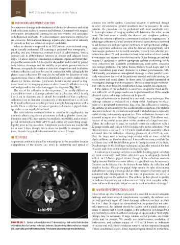
wall and gas within the collection suggest the diagnosis (Fig. 30-1).
The size of the collection is also important. It is usually difficult or If the nature of the collection is uncertain, diagnostic fluid aspira-
impossible to insert a drainage catheter into a collection, which is only tion with a 20- or 22-gauge needle can be performed first. If the sample
1 or 2 cm in diameter, and it should be remembered that a spherical obtained is pus, a drainage catheter can be placed.
Large collections can be drained by a one-stick, trocar technique. The
collection 2 cm in diameter contains only a little more than 4 cc of fluid.
With small collections we often perform a simple fluid aspiration with a drainage catheter is preloaded on a sharp stylet. Analogous to place-
ment of a peripheral intravenous line, once the collection is entered,
needle. Once a collection is 3 cm or greater in diameter, a pigtail drain-
age catheter can usually be secured. the catheter is advanced over the needle into the collection. The stylet is
then removed and the contents are aspirated. This technique is especially
The main relative contraindication to consider is coagulopathy. We
routinely obtain coagulation parameters including platelet count, pro- useful during endocavitary approaches. Most collections, however, are
accessed using an over-the-wire Seldinger technique. This allows veri-
thrombin time (PT), international normalized ratio (INR), and activated
partial thromboplastin time (aPTT) and correct any underlying coagu- fication of successful access prior to the creation of a large bore tract.
Unless the collection is large, we typically enter the collection with a
lopathy prior to the procedure. Antiplatelet medications are ideally held
for at least 3 days, though this is often not feasible in emergent situa- 22-gauge needle and coil an 0.018-in guide wire in the collection. Over
this microwire, a coaxial 5- or 6-French sheath/dilator assembly is then
tions. Heparin is typically discontinued for at least 2 hours. advanced into the collection, allowing placement of a 0.035-in wire.
■ TECHNIQUE Over the larger wire, a locking loop catheter with an inner metal or
Appropriate antibiotics should be initiated prior to the procedure because plastic stiffener can then be advanced. It is usually necessary to dilate the
soft tissue tract with fascial dilators prior to final placement of the drain.
manipulation of the abscess can result in bacteremia and spread of Disadvantages of the Seldinger technique include the potential for loss
of access and cross-contamination during exchanges.
A wide array of drainage catheters is available. Locking pigtail catheters
are most commonly used. Most collections can be adequately drained
with 6- to 12-French pigtail drains, though if the collection contains
highly viscous fluid or extensive debris, a larger drain may be necessary.
Contrast can be injected into the drain to better define the collection and
visualize fistulas. Though the pigtail helps secure the tube, skin sutures
and adhesive locking dressings add an extra measure of security against
accidental tube dislodgement. At the time of placement, we strive to
completely aspirate the collection. The catheter is then placed to gravity
or bulb suction and output is documented. With thick complex collec-
tions, saline or fibrinolytic irrigation can be used to facilitate drainage. 3
■ IMMEDIATE POSTPROCEDURAL CARE
Close follow-up after catheter placement is essential to ensure adequate
drainage and detect delayed complications. Normally, the catheter out-
put will gradually taper off. Most drainage catheters are kept in place
for 3 to 7 days. If output has diminished but the patient has not clini-
cally improved, the catheter should be flushed with a small amount of
saline to ensure that it is not clogged. If the catheter is not clogged but
appropriately positioned, catheter exchange or upsize and/or fibrinolytic
therapy may be necessary. If large volume output persists, an enteric
fistula may be present. We usually use defervescence, resolution of
FIGURE 30-1. Contrast-enhanced abdominal CT demonstrating a thick-walled fluid collection leukocytosis, and catheter output of <10 cc/24 hours as indicators
with multiple foci of air (arrows) in the right abdomen. The patient was febrile and had an elevated of success and will consider catheter removal without repeated imaging
WBC count status post right hemicolectomy. Percutaneous abscess drainage revealed frank pus. if these conditions are met. If not, repeat imaging should be performed.
section02.indd 210 1/13/2015 2:05:47 PM