
Page 309 - Hall et al (2015) Principles of Critical Care-McGraw-Hill
P. 309
CHAPTER 30: Interventional Radiology 213
patients, a bridge to adjunctive therapies such as gallbladder ablation, fluoroscopic guidance is used for tract dilatation and catheter place-
stone dissolution, shock-wave lithotripsy, and/or basket extraction. 13-16 ment. In trocar technique, a small-bore catheter fitted over a stiffening
Adjunctive techniques for stone removal have been associated with a cannula and sharp stylet is advanced as a unit under US guidance, and
high rate of gallstone recurrence in retrospective studies—10% to 30% the catheter is advanced off the cannula directly into the gallbladder.
per year with a symptomatic recurrence rate of approximately 6% to Most radiologists place self-retaining, locking loop catheters. While
18% per year. Therefore, most high-risk patients undergo eventual there is likely no difference in the incidence of peritonitis after trans-
17
surgical cholecystectomy, and poor candidates may require permanent peritoneal versus transhepatic placement, transhepatic placement may
cholecystostomy. improve stability during and after placement and is favored by some
In the case of acute acalculous cholecystitis, the drainage catheter can radiologists. Gram stain and culture results of the bile are not sensi-
be removed after resolution in most cases, without the need for elec- tive (30%-50%) but may aid in determining specific antibiotic therapy
tive interval cholecystectomy, since the risk of recurrence is likely to be when positive.
low (<10%) based on retrospective studies. Predisposing conditions
15
include diabetes, malignancy, burn injury, recent surgery, recent trauma, ■ POSTPROCEDURE CARE
cardiac disease, positive pressure ventilation, and total parenteral nutri-
tion. Establishing the diagnosis remains a clinical challenge since the Cholecystostomy catheters are drained to gravity bag, and output is
accuracy of US (Fig. 30-6) is approximately 50% to 60%, and the false- monitored every shift. If the cystic duct is indeed obstructed, low
positive rate of nuclear medicine hepatobiliary scans is approximately volumes of clear mucus (50-70 mL) are expected daily. Larger volumes
25% to 30%, caused by factors such as liver dysfunction, sepsis, fasting, of biliary drainage indicate cystic duct patency, and very large volumes
and prolonged total parenteral nutrition. In many cases, a high clinical (>1 L) indicate obstruction of the distal common bile duct and patency
suspicion by the critical care team leads to PC in the setting of soft radio- of the cystic duct, usually the result of stone migration. Management of
logical support. Patients with true acute acalculous cholecystitis typically the cholecystostomy catheter is typically a combined effort by the ICU
show a quick and marked clinical response to PC. team and IR staff. New onset bleeding, leakage of bile around the skin
■ TECHNIQUE tube dislodgment or obstruction, and evaluation under fluoroscopy or
entry site, or lack of timely resolution of clinical symptoms may indicate
PC is best performed in the IR suite using US and fluoroscopic guidance by cross-sectional imaging may be indicated.
The need for prolonged catheterization should be managed with fluo-
under moderate sedation, but can be done in select cases at the bedside roscopically guided catheter changes every 4 to 6 weeks. After clinical
using only portable US guidance. Difficult or complicated cases may resolution, patients with acalculous cholecystitis may undergo contrast
require CT guidance. injection under fluoroscopy. The criteria for catheter removal include
Patients are typically referred to interventional radiology after the absence of gallstones, patency of the cystic and common bile ducts,
initiation of broad-spectrum antibiotic coverage; otherwise, preproce- free spillage of contrast into the duodenum (Fig. 30-8), and the verifi-
dural antibiotics are administered. Authors have described successful cation of a mature tract by over-the-wire contrast injection, typically
applications of both Seldinger and trocar techniques. The advantage of present at 4 to 6 weeks (Fig. 30-9). Patients with calculous cholecys-
Seldinger technique is verification of creation of an access tract to the titis face the options of surgical cholecystectomy, adjunctive therapies
gallbladder using a low-gauge needle prior to dilation and placement of described above, or permanent cholecystostomy.
a drainage catheter. The advantage of trocar technique is placement of a
associated with serial tract dilatation. In Seldinger technique, a 21- or ■ RESULTS AND COMPLICATIONS
drainage catheter in a single step, without the potential for bile leakage
22-gauge needle is advanced into the gallbladder under US guidance, Technical success exceeds 95%. Clinical success is complicated by
19
bile is aspirated, contrast is gingerly injected (Fig. 30-7), a coaxial the absence of true cholecystitis in many cases, but is approximately
dilator is used to convert from microwire to standard wire access, and 60% for patients with suggestive US findings. Major periprocedural
20
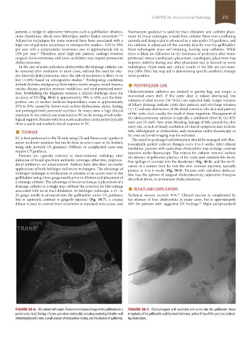
FIGURE 30-6. ICU patient with sepsis. Transverse ultrasound image of the gallbladder in a FIGURE 30-7. Cholecystogram with successful wire access into the gallbladder shows
patient with classic findings of acute acalculous cholecystitis, including marked gallbladder wall irregularity of the gallbladder wall, luminal distension, and no filling of the cystic duct indicat-
thickening beyond 3 mm, a small amount of intraluminal sludge, and the absence of gallstones. ing obstruction.
section02.indd 213 1/13/2015 2:05:49 PM