
Page 307 - Hall et al (2015) Principles of Critical Care-McGraw-Hill
P. 307
CHAPTER 30: Interventional Radiology 211
■ RESULTS AND COMPLICATIONS
Technical success exceeds 90% and is immediately apparent. Clinical
success rates depend on abscess location and complexity, underlying
immune status, and subsequent fistula formation. Overall, however,
clinical success ranges from 70% to 90%. Overall complication rates
for percutaneous abscess drainage range from 10% to 15%, with seri-
ous complications accounting for less than 5% of cases. Infectious
4
complications may be encountered during primary catheter placement,
when spread of abscess contents or bacteremia can occur. As a result of
prolonged catheterization, skin-site infections can occur. Hemorrhage
can occur from venous or arterial transection or from the development
of pseudoaneurysms or vascular fistulas. Minor bleeding is usually
self-limited and managed conservatively. In these cases, temporary tube
capping, upsizing, or repositioning may help tamponade the bleeding.
When more substantial hemorrhage is suspected, angiography and sur-
gical consultation are usually necessary. Inadvertent catheterization or
puncture of adjacent organs or bowel may also occur.
PERCUTANEOUS NEPHROSTOMY
KEY POINTS
• Percutaneous nephrostomy should be performed for urinary diver-
sion when retrograde ureteral stenting fails or is contraindicated.
• Hematuria is very common after nephrostomy and should slowly
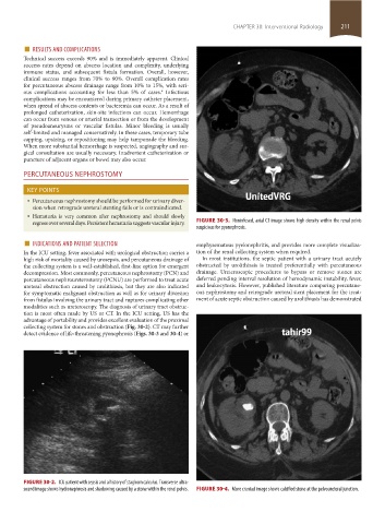
regress over several days. Persistent hematuria suggests vascular injury. FIGURE 30-3. Noninfused, axial CT image shows high density within the renal pelvis
suspicious for pyonephrosis.
■ INDICATIONS AND PATIENT SELECTION emphysematous pyelonephritis, and provides more complete visualiza-
In the ICU setting, fever associated with urological obstruction carries a tion of the renal collecting system when required.
high risk of mortality caused by urosepsis, and percutaneous drainage of In most institutions, the septic patient with a urinary tract acutely
the collecting system is a well-established, first-line option for emergent obstructed by urolithiasis is treated preferentially with percutaneous
decompression. Most commonly, percutaneous nephrostomy (PCN) and drainage. Ureteroscopic procedures to bypass or remove stones are
percutaneous nephroureterostomy (PCNU) are performed to treat acute deferred pending interval resolution of hemodynamic instability, fever,
ureteral obstruction caused by urolithiasis, but they are also indicated and leukocytosis. However, published literature comparing percutane-
for symptomatic malignant obstruction as well as for urinary diversion ous nephrostomy and retrograde ureteral stent placement for the treat-
from fistulas involving the urinary tract and ruptures complicating other ment of acute septic obstruction caused by urolithiasis has demonstrated
modalities such as ureteroscopy. The diagnosis of urinary tract obstruc-
tion is most often made by US or CT. In the ICU setting, US has the
advantage of portability and provides excellent evaluation of the proximal
collecting system for stones and obstruction (Fig. 30-2). CT may further
detect evidence of life-threatening pyonephrosis (Figs. 30-3 and 30-4) or
FIGURE 30-2. ICU patient with sepsis and a history of staghorn calculus. Transverse ultra-
sound image shows hydronephrosis and shadowing caused by a stone within the renal pelvis. FIGURE 30-4. More craniad image shows calcified stone at the pelvoureteral junction.
section02.indd 211 1/13/2015 2:05:48 PM