
Page 311 - Hall et al (2015) Principles of Critical Care-McGraw-Hill
P. 311
CHAPTER 30: Interventional Radiology 215
been used successfully though medium-sized particles are most commonly entirely account for the hemoptysis, a search of nonbronchial systemic col-
used. The angiogram should be carefully evaluated for potential bronchial lateral supply is performed (Fig. 30-10C).
then enter the systemic arterial circulation. If these shunts are present, larger ■ IMMEDIATE POSTPROCEDURAL CARE
artery to pulmonary vein shunts. Smaller particles could traverse shunts and
particles or coils may be necessary to close the shunt prior to proceeding The patient must lie flat for 2 to 6 hours depending on whether or not an
with the embolization. If the bronchial arteries appear normal or do not arterial closure device was deployed. Special attention should be paid to
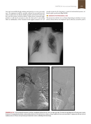
FIGURE 30-10. A. Chest radiograph demonstrates multifocal consolidation and bronchiectasis, worst in the right upper lobe. The patient had multidrug resistant TB and recurrent massive
hemoptysis. B. Angiography demonstrates an intercostobronchial trunk. The bronchial artery is tortuous and hypertrophied with dense parenchymal blush. C. Angiography of the right internal
mammary artery (arrowhead) shows pulmonary parenchymal blush (arrow) contributing to the hemorrhage.
section02.indd 215 1/13/2015 2:05:54 PM