
Page 310 - Hall et al (2015) Principles of Critical Care-McGraw-Hill
P. 310
214 PART 2: General Management of the Patient
BRONCHIAL ARTERY EMBOLIZATION
KEY POINTS
• Most clinically significant episodes of hemoptysis are caused by
bleeding from the bronchial arteries.
• Chronic pulmonary diseases (eg, cystic fibrosis, tuberculosis)
are the most common underlying disorders predisposing to life-
threatening hemoptysis.
• Bronchial artery embolization is an effective and safe treatment of
massive hemoptysis.
Massive hemoptysis, defined as bleeding greater than 300 mL/24 hours,
carries a mortality rate of up to 85% in patients treated by conservative
means. Recurrent bouts of moderate hemorrhage are also life threaten-
21
ing. Death is usually due to asphyxiation rather than exsanguination or
hemorrhagic shock. Surgical resection can be curative for focal disease, but
it carries a high mortality rate in the setting of acute hemorrhage. Bronchial
artery embolization has proven to be an effective and safe treatment. 22,23
■ INDICATIONS AND PATIENT SELECTION
Massive hemoptysis typically occurs in the setting of chronic inflam-
matory lung disease. In 90% of patients, bleeding predominantly arises
from the bronchial artery. Tuberculosis, sarcoidosis, and cystic fibro-
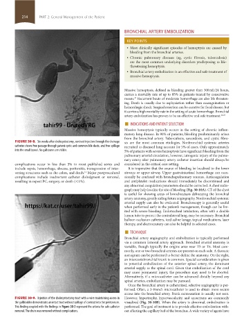
FIGURE 30-8. Six weeks after cholecystostomy, contrast injection through the drainage sis are the most common etiologies. Nonbronchial systemic arteries
catheter shows free passage through patent cystic and common bile ducts, and free spillage recruited to diseased lung account for 5% of cases. Only approximately
into the small bowel. No gallstones are visible. 5% of patients with severe hemoptysis have significant bleeding from the
pulmonary arterial circulation, however, iatrogenic injury of the pulmo-
nary artery after pulmonary artery catheter insertion should always be
complications occur in less than 5% in most published series and considered in the critical care setting.
include sepsis, hemorrhage, abscess, peritonitis, transgression of inter- It is imperative that the source of bleeding be localized to the lower
vening structures such as the colon, and death. Major postprocedural airways or upper airway. Upper gastrointestinal hemorrhage can occa-
11
complications include inadvertent catheter dislodgment or removal, sionally be confused with bronchopulmonary sources. Anticoagulation
resulting in repeat PC, surgery, or death (<1%). and antiplatelet medications should immediately be discontinued and
any abnormal coagulation parameters should be corrected. A chest radio-
graph may help localize the site of bleeding (Fig. 30-10A). CT of the chest
is useful for showing areas of bronchiectasis delineating the bronchial
artery anatomy, greatly aiding future angiography. Nonbronchial systemic
arterial supply can also be evaluated. Bronchoscopy is generally useful
when performed early in the patient’s management, though can be lim-
ited with severe bleeding. Endotracheal intubation, often with a double
lumen tube to protect the contralateral lung, may be necessary. Bronchial
balloon occlusion catheters, iced saline lavage, topical medications, laser
therapy, and electrocautery can also be helpful in selected cases.
■ TECHNIQUE
Bronchial artery angiography and embolization is typically performed
via a common femoral artery approach. Bronchial arterial anatomy is
variable, though typically the origins arise near T5 or T6. Most com-
monly, one or two bronchial arteries are present on each side. A thoracic
aortogram can be performed to better define the anatomy. On the right,
an intercostobronchial trunk is common. Special consideration is given
to potential embolization of the anterior spinal artery, the dominant
arterial supply to the spinal cord. Given that embolization of the cord
may cause permanent injury, the procedure may need to be aborted.
Alternatively, if a microcatheter can be advanced distally beyond any
spinal arteries, embolization may be pursued.
Once the bronchial artery is catheterized, selective angiography is per-
formed. Often, a 3-French microcatheter is used to obtain more secure
access into the bronchial artery. Frank extravasation is usually not seen.
FIGURE 30-9. Injection of the cholecystostomy tract with a wire maintaining access in However, hypertrophy, hypervascularity, and aneurysms are commonly
the gallbladder demonstrates an intact tract without spillage of contrast into the peritoneum. visualized (Fig. 30-10B). When the artery is abnormal, embolization is
This finding coupled with the findings in Figure 30-3 represent the criteria for safe catheter performed. The goal of treatment is to provide effective embolization with-
removal. The drain was removed without complications. out affecting the capillary bed of the bronchus. A wide variety of agents have
section02.indd 214 1/13/2015 2:05:50 PM