
Page 308 - Hall et al (2015) Principles of Critical Care-McGraw-Hill
P. 308
212 PART 2: General Management of the Patient
similar technical and clinical success rates, and both procedures percutaneous nephrolithotomy by basket or snare in the IR suite, or
5,6
remain viable, first-line options for this indication. The presence of require extracorporeal shock wave lithotripsy by urologists. If prolonged
thrombocytopenia associated with sepsis makes percutaneous access PCN or PCNU is required, fluoroscopically guided catheter exchange
less optimal in many cases, and severe, uncorrectable coagulopathy is is recommended every 4 to 6 weeks. PCNU catheters can be capped
the main, relative contraindication to PCN. 7 for patient comfort if the patient remains asymptomatic. In many cases
■ TECHNIQUE involving neoplastic, fibrotic, inflammatory, or iatrogenic obstruction,
prolonged internal drainage is preferred to external drainage for patient
PCN and PCNU are best performed in the IR suite using a combina- comfort, and the PCN or PCNU can be converted to internal double J
tion of US and fluoroscopic guidance with the patient in the prone or ureteral stents in the IR suite. Internal stents require routine cystoscopi-
semiprone position under moderate sedation. Preprocedural antibiotics cally guided changes every 3 months by urology.
initiated prior to referral to radiology. Using Seldinger technique, a 21- ■ CATHETER MANAGEMENT
are administered unless broad-spectrum antibiotic coverage has been
or 22-gauge needle is advanced through a posterior calyx into the renal Management of the PCN catheter is typically a combined effort by the
pelvis under US guidance, urine is aspirated to verify access, contrast ICU team and rounding IR staff. Typically, the collected urine progresses
is gingerly injected, a coaxial dilator is used to convert from microwire from blood tinged to clear over 2 to 3 days. Severe bleeding at the time of
to standard wire access, and fluoroscopic guidance is used during tract placement may respond to capping the catheter for a few hours to create
dilatation and catheter placement. Performance of a diagnostic neph- a tamponade effect. Delayed onset of bleeding or persistent low-grade
rostogram involves intraluminal distension by contrast material with the bleeding is typically caused by venous injury and addressed by reposi-
risk of symptom exacerbation, and is often deferred pending resolution tioning or upsizing the catheter under fluoroscopic guidance. Leakage of
of fever and leukocytosis. The retention mechanism of a self-retaining urine around the skin entry site, or lack of timely resolution of clinical
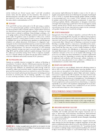
catheter (typically locking-loop type) is secured within the renal pelvis symptoms, may indicate tube dislodgment or obstruction, and evalua-
(Fig. 30-5), and the catheter is placed to gravity drainage. Urine is sent tion under fluoroscopy or by cross-sectional imaging may be indicated.
for culture and sensitivity testing. When PCNU is required, manipula- Inadvertent retraction of the tube can be treated with exchange, if any
tion of a 5-French catheter and guide wire into the urinary bladder pre- access into the kidney has been maintained, or complete replacement
cedes tract dilatation and internal-external PCNU placement. if access has been lost. Depending on severity, skin-site infections can
■ POSTPROCEDURE CARE be addressed with antibiotics and fluoroscopic catheter evaluation, or
in more severe cases, placement of a new catheter at a different site or
Patients are carefully observed overnight for evidence of bleeding or internalization to a double J stent if this option exists.
the kidney. Fluid input and catheter output are recorded every shift, and ■ RESULTS AND COMPLICATIONS
exacerbation of systemic infection following percutaneous drainage of
broad-spectrum antibiotics are administered until coverage needs are PCN is successful in cases of dilated, obstructed collecting systems in
dictated by culture and sensitivity results. Catheters typically remain in 98% to 99% of cases in published literature spanning decades. Lower
7
place until fever and leukocytosis have resolved, the cause of the obstruc- success rates are encountered in the absence of pelvocaliectasis and in
tion has been treated, and adequate time has passed for healing and tract the presence of complex staghorn calculi. Major and minor complica-
formation to minimize the risk of bleeding—typically 1 to 2 weeks. tions occur in approximately 10% of patients. The most common major
In cases involving urolithiasis, stones may pass spontaneously, require complication is sepsis or exacerbation of systemic infection, most com-
monly associated with the presence of pyonephrosis. Overdistension of
8
the renal collecting system should be strictly avoided in these patients.
Pleural complications such as pneumothorax, hemothorax, or empyema
reportedly occur in 9% to 12% of patients undergoing PCN via an inter-
costal window. Other reported major complications are less common
9
and include hemorrhage and colon transgression.
PERCUTANEOUS CHOLECYSTOSTOMY
KEY POINTS
• Indications for percutaneous cholecystostomy appear to be increasing.
• Cholecystostomy catheters must remain in place long enough (usu-
ally >2 weeks) for a track to mature prior to manipulation or removal.
■ INDICATIONS AND PATIENT SELECTION
Patients with acute cholecystitis in the ICU are often at high risk for
morbidity and mortality associated with surgical treatments such as
open or laparoscopic cholecystectomy. Percutaneous cholecystostomy
(PC) has been established as a definitive treatment, a bridge to surgery,
or a means toward adjunctive, minimally invasive therapies, depending
on patient presentation. 10,11
In the case of acute calculous cholecystitis, surgical cholecystec-
tomy remains the first-line therapy in surgical candidates. In low-risk
patients, published periprocedural mortality rates of both open and lap-
12
FIGURE 30-5. A locking-loop pigtail nephrostomy catheter has been placed in the renal aroscopic cholecystectomy are typically below 1%. In patients deemed
pelvis. Minimal contrast injection shows limited filling of the pelvicalyceal system secondary too unstable to undergo surgery and/or general anesthesia, PC serves as
to a combination of intraluminal stones and pus. bridge to more elective surgery or, in permanently high-risk, comorbid
section02.indd 212 1/13/2015 2:05:48 PM