
Page 361 - Hall et al (2015) Principles of Critical Care-McGraw-Hill
P. 361
CHAPTER 31: The Pathophysiology of the Circulation in Critical Illness 231
such as Pla are measured with respect to atmospheric pressure, so they decreased VR by increasing Pra, thus keeping end-diastolic volume and
do not represent true transmural, or filling, pressures of the heart cham- Q ˙ t abnormally low. Tension pneumothorax, massive pleural effusions,
ber when the pressure on the outside of the heart is not atmospheric. high levels of PEEP, and greatly increased abdominal pressures can
5,6
Pericardial pressure is most often equal to Ppl, which is subatmospheric increase pressure outside the heart (Ppl) and thus reduce LVEDV and
during spontaneous breathing (−3 to −10 mm Hg) and can become SV despite high values of LVEDP (Table 31-1). Intercurrent LV hyper-
very negative in airflow obstruction or very positive with mechanical trophy or infiltrative diseases (amyloidosis) occasionally stiffen the
ventilation and positive end-expiratory pressure (PEEP). For conve- relaxed ventricle such that high filling pressures are needed to maintain
nience, the following discussion refers to the intravascular pressures as an adequate SV, and inadequate filling time or poorly coordinated atrial
transmural, or filling, pressures, and any cause for altered pericardial or contraction also impairs ventricular filling. 7
pleural pressure is noted. A right-to-left shift of the interventricular septum can also restrict
■ THE DIASTOLIC V-P CURVE AND VENTRICULAR diastolic filling. Presumably, the distention of the right ventricle causes
the interventricular septum to bulge from right to left, thereby reduc-
FILLING DISORDERS (SEE TABLE 31-1) ing the unstressed volume and compliance of the left ventricle. This
2,8
Figure 31-4B plots LVEDV against LVEDP. As ventricular volume effect of ventricular interdependence is much less marked when the
increases from zero, the transmural pressure of the ventricle does not pericardium is removed, perhaps because the limiting membrane of
exceed zero until about 50 mL (the unstressed volume) is added. Then the pericardium restricts freedom of motion of the left ventricle, mak-
LVEDP increases in a curvilinear manner with ventricular volume (the ing it more vulnerable to displacements of the septum. Accordingly,
stressed volume) first as a large change in volume for a small change conditions in which the right ventricle is abnormally loaded (eg, acute
in pressure and then as a small change in volume for a large change in pulmonary embolism or acute-on-chronic respiratory failure due to
pressure. If the pericardium is removed, these V-P characteristics are obstructive or restrictive lung disease) may impede the emptying of the
more linear such that the large change in LVEDP at higher values of right ventricle, causing it to work at a higher end-diastolic volume. Then
LVEDV is no longer evident. Thus the pericardium acts like a mem- LV filling pressures will be higher than expected for the end-diastolic
brane with a large unstressed volume loosely surrounding the heart up volume. This provides one possible explanation for why PEEP is often
to a given ventricular volume, but at greater LVEDV the pericardium associated with increased filling pressure to maintain a normal SV even
becomes very stiff. At higher heart volumes, most of the pressure when LVEDP is corrected to the true filling pressure by subtracting
5,6,9,10
across the heart is across the pericardium, accounting for the very the increase in Ppl (ΔPpl) measured when PEEP is applied. Acute
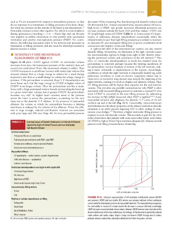
steep rise in the diastolic V-P relation. In the presence of pericardial myocardial ischemia also displaces the diastolic V-P curve of the left
effusion, the volume at which the pericardium becomes a limiting ventricle up and to the left (Fig. 31-5). Conceivably, myocardial injury
membrane is reduced by the volume of the effusion. When the effu- and ischemia alter the elastic properties of the relaxed ventricle as diastolic
sion is large enough, reduced end-diastolic volumes are associated relaxation is an active process requiring ATP to allow cycling of actin-
6,11
with quite large end-DPs (see Chap. 40). In turn, pericardial pressure myosin cross-bridges. Therefore, a higher ventricular filling pressure is
required at each end-diastolic volume. This accounts in part for the often
noted observation that patients with acute myocardial injury need values
TABLE 31-1 Common Causes of Diastolic Dysfunction in Critically Ill Patients of LVEDP as high as 30 mm Hg to maintain adequate Q ˙ t, whereas normal
Signaled by High Left Atrial Pressure and Low Ventricular patients need filling pressures below 10 mm Hg.
End-Diastolic Volume
External compression
Pericardial effusion or constriction
Positive pressure ventilation with PEEP, auto-PEEP
Tension pneumothorax, massive pleural effusions
100
Greatly increased abdominal pressure
Myocardial stiffness
LV hypertrophy—aortic stenosis, systemic hypertension
Infiltrative diseases—amyloidosis Left ventricular pressure
Ischemic heart disease
Ventricular interdependence and right-to-left septal shift 50
Pulmonary hypertension
RV infarction
High levels of PEEP
Severe acute hypoxic respiratory failure
Intraventricular filling defects
0 50 100 150
Tumor
Left ventricular volume
Clot
FIGURE 31-5. Schematic representation of left ventricular end-diastolic volume (LDEDV)
Rhythm or valvular impediments to filling
and pressure (LVEDP) and end-systolic (ES) volume and pressure relations before (continuous
Tachycardia curves) and after (interrupted curves) acute myocardial infarction. The myocardial injury depresses
Heart block the contractility to increase ES volume despite the decrease in pressure afterload; accordingly,
LVEDV increases to accommodate venous return, whereas LVEDP increases even more due to the
Atrial fibrillation, flutter
diastolic dysfunction of the myocardial injury. Accordingly, LV dysfunction is signaled by reduced
Mitral stenosis stroke volume and cardiac output despite a large elevation in LVEDP; therapy aims to reduce
LV, left ventricular; PEEP, positive end-expiratory pressure; RV, right ventricular. preload, enhance contractility, and reduce afterload. For further discussion, see text.
section03.indd 231 1/23/2015 2:06:40 PM