
Page 366 - Hall et al (2015) Principles of Critical Care-McGraw-Hill
P. 366
236 PART 3: Cardiovascular Disorders
increased, and although Q ˙ t usually increases further with intravenous
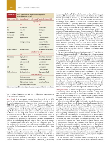
TABLE 31-2 Initial Approach to the Diagnosis and Management
of the Hypotensive Patient infusions, BP increases little with increased Q ˙ t. Further, the need for
an even greater Q ˙ t to increase D is questionable because the lactic
˙
Blood Pressure (BP) = Cardiac Output (Qt) × Systematic Vascular Resistance (SVR) acidosis of septic shock may not be due to anaerobic metabolism. 32-34
O 2
˙
Is Qt Reduced? Accordingly, septic patients in whom Q ˙ t is maximized do not have
improved survival. 35,36 Conversely, pulmonary vascular pressures always
Yes No increase with volume infusion, thus increasing pulmonary edema when
BP 90/70 mm Hg 90/40 mm Hg the septic process increases the permeability of lung vessels. 31,37-39 This
coincidence of the acute respiratory distress syndrome (ARDS) and
Skin Cool, blue Warm, pink
septic shock has created an apparent dilemma concerning fluid therapy
Nail bed return Slow Rapid and cardiovascular management of these conditions. One approach is
40
Heart sounds Muffled Crisp to ensure resuscitation from septic shock as the first priority by ensur-
ing a large Q ˙ t with a Ppw that does not exceed 15 mm Hg or a CVP of
History/lab Hypervolemic or ↓ or ↑ WBC and/or
temperature 8 to 12 mm Hg in the absence of pulmonary catheter measurements
and add dobutamine to increase Q ˙ t and BP as necessary. As noted
31
Cardiogenic etiology Source of infection above, however, optimal Q ˙ t does not equal maximal Q ˙ t 41,42 and the
Immune compromise benefit from an increased Q ˙ t in response to inotropic agents needs to
43
Severe liver disease be weighed against the risk of tachydysrhythmias. When early ARDS is
not associated with septic shock, we seek the lowest circulating volume
Working diagnosis See next question Septic shock/endotoxemia
to provide adequate Q ˙ t. 31
Is the Heart Too Full? The septic myocardium does not function normally, 44,45 but this dys-
Yes No function is often associated with SV values larger than 100 mL at normal
values of LVEDP. Accordingly, it seems unlikely that systolic dysfunc-
Presentation Angina, dyspnea Hemorrhage, dehydration
tion contributes substantially to the shock, but infusion of dobutamine
Signs Cardiomegaly Dry mucous membranes does increase Q ˙ t for a given high-normal LVEDP without increasing
41
Extra heart sounds ↓ tissue turgor O uptake or correcting lactic acidosis in septic shock. Even when Q ˙ t
2
and D are made adequate with fluid and dobutamine infusions, the
↑ JVP Stool, gastric blood O 2
perfusion pressure for vital organs such as the brain and heart may still
Lab ECG, x-ray ↓ hematocrit be too low in some septic patients. In this case, norepinephrine infu-
Echocardiogram ↑ BUN/creatinine sion increases BP and splanchnic blood flow 46,47 without compromising
renal function ; in contrast, dopamine and epinephrine infusions cause
48
Working diagnosis Cardiogenic shock Hypovolemic shock
splanchnic hypoperfusion in septic shock and due to their β effects are
1
What Does Not Fit? also associated with tachydysrhythmias. 46-50 Tachypnea and respiratory
Cardiac tamponade Anaphylaxis distress may be severe, so initial supportive therapy includes consider-
Acute pulmonary hypertension Spinal shock ation of early intubation and mechanical ventilation and correction of
hyperthermia with antipyretics, paralysis, and cooling. This prevents
Right ventricular infarction Adrenal insufficiency catastrophic respiratory muscle fatigue, respiratory acidosis, and the
Overlapping multiple etiologies complications of emergent intubation and may improve tissue oxygen-
BUN, serum urea nitrogen; ECG, electrocardiogram; JVP, jugular venous pressure; WBC, white blood cell count. ation by reducing O requirements in patients with limited D . 51,52
2
O 2
Cardiogenic Shock: In contrast to septic shock, low Q ˙ t is signaled by low
PP indicating low SV (see Fig. 31-3), signs of increased SVR (eg, cold,
history, physical examination, and routine laboratory tests to answer blue, damp extremities and poor return of color to the nail bed), and
three questions in sequence. a history or presentation including features suggesting a cardiogenic
or hypovolemic cause of hypotension. If Q ˙ t is reduced in the hypoten-
Septic Shock: Is BP decreased because Q ˙ t is decreased? If not, SVR sive patient, then the heart may be too full.
must be reduced, a condition almost always related to sepsis or ster- A heart that is too full (see Table 31-2) is often signaled by symptoms
ile endotoxemia associated with severe liver disease. As indicated in of ischemic heart disease or arrhythmia, signs of cardiomegaly, the third
Table 31-2 (right column), a low BP is often characterized by a large and fourth sounds or gallop rhythm of heart failure, new murmurs of
PP because the SV is large and by a very low DP because each SV has valvular dysfunction, increased jugular or CVP, and laboratory tests
a rapid peripheral runoff through dilated peripheral arterioles (see suggesting ischemia (eg, electrocardiogram [ECG], creatine phosphoki-
Fig. 31-3). This produces warm, pink skin with rapid return of color nase, or troponin determination) or ventricular dysfunction (eg, chest
to the nail bed and crisp heart sounds. As in other types of shock, x-ray suggesting cardiomegaly, a widened vascular pedicle, or cardio-
tachycardia is evident due in part to baroreceptor reflex response genic edema or echocardiogram showing regional or global systolic
to hypotension, but the arterial vasoconstriction response to reflex dyskinesia). The most common cause of hypotension associated with a
sympathetic tone is blocked by relaxation of arteriolar smooth muscle circulation that is too full on initial evaluation is cardiogenic shock due
induced by endothelium-derived relaxing factor (or nitric oxide). to myocardial ischemia (see Chaps. 35 and 37). Initial therapy treats
The combination of tachycardia and large PP indicates a large Q ˙ t this presumptive diagnosis with inotropic drug therapy (dobutamine
that is almost always present early unless concurrent hypovolemia or 3-10 µg/kg per minute) to assist the ejecting function of the ischemic
myocardial dysfunction precludes the hyperdynamic circulatory state heart. Such therapy does not directly address the coronary insufficiency
of sepsis. and may increase the myocardial O demand, especially if it causes
2
Initial therapy starts with appropriate broad-spectrum antibiotics (see tachycardia. Concurrent sublingual, dermal, or intravenous nitroglyc-
Chap. 64) and includes expansion of the circulating volume by intrave- erin ameliorates elements of coronary vasospasm to increase blood flow
nous infusion of fluids to treat associated hypovolemia, which is due to and reduces preload to decrease myocardial O consumption. Morphine
2
venodilation decreasing Pms and VR lower than needed to maintain also decreases pain, anxiety, and preload. 53
adequate perfusion pressure of vital organs. The end point of volume In this situation, even a cautious volume challenge may be risky
infusion is obscure because Q ˙ t and oxygen delivery (D ) are already because ventricular function and Q ˙ t are decreased as often as they are
O 2
section03.indd 236 1/23/2015 2:06:43 PM