
Page 384 - Hall et al (2015) Principles of Critical Care-McGraw-Hill
P. 384
254 PART 3: Cardiovascular Disorders
■ CATHETERS AND MONITORING DURING INITIAL RESUSCITATION 150
After an airway is established and breathing ensured, correction of the
circulatory abnormality always requires good intravenous access. For
large-volume administration, two peripheral intravenous catheters of
gauge 16 or larger or large-bore central venous access is required. Early 100
Goal-Directed Therapy mandates immediate placement of a central
venous catheter. Electrocardiographic monitoring is easily accomplished
and usefully measures heart rate and rhythm for early detection and, LV Pressure
hence, rational treatment of tachyarrhythmias or bradyarrhythmias
aggravating the low-flow state. 50
The urinary bladder should be catheterized to measure urine output
and to facilitate urine sampling. A nasogastric or orogastric tube to
decompress the stomach and later to deliver medication and nutrition is
generally required in the intubated patient. Measuring arterial pressure
with a peripheral arterial or femoral arterial catheter is useful because, in 0
the patient in shock with low cardiac output or low blood pressure, cuff 0 50 100 150
pressures may be inaccurate. Appreciation of MAP, pulse pressure (related LV Volume
1
to stroke volume), and pulse pressure variation with respiration (values
greater than ~15% suggest volume-responsive hypovolemia ) are enabled.
26
Arterial blood-gas and other blood samples are also readily obtained. 12
Effective use and interpretation of Scv and echocardiography often
O 2
obviate pulmonary artery catheterization when the clinical hypothesis
of hypovolemic, cardiogenic, or septic shock is confirmed and corrected 10
by initial therapeutic intervention. There is no role for pulmonary artery
catheterization for routine monitoring or management of uncompli- 8
cated shock states. 27,28 Use of pulmonary artery catheterization should be
restricted to circumstances in which the derived measurements will alter
management or direct therapeutic interventions. Cardiac output 6 Normal
■ TEMPO 4
One of the most important contributions the intensivist can make to the Hypovolemic Shock
care of a shock patient is to establish an appropriately rapid management 2
tempo. Rapid initial resuscitation improves survival (“time is tissue”). In
many instances, resuscitation driven by protocol can achieve adequate 0
resuscitation faster. Effective protocol-driven resuscitation requires –5 0 5 10 15 20 25
significant preliminary discussion, buy in, and training of emergency End-diastolic pressure
room physicians, house staff, nurses, respiratory therapists, and others.
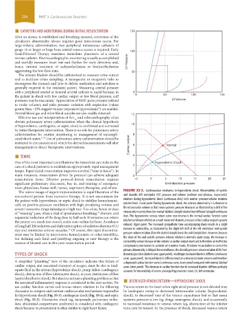
The mirror image of urgent implementation is rapid liberation of the FIGURE 33-3. Cardiovascular mechanics in hypovolemic shock. Abnormalities of systolic
resuscitated patient from excessive therapy. It is not uncommon for and diastolic left ventricular (LV) pressure and volume (ordinate and abscissa, respectively)
the patient with hypovolemic or septic shock to stabilize hemodynami- relations during hypovolemic shock (continuous lines) with normal pressure-volume relations
cally on positive-pressure ventilation with high circulating volume and (dashed lines). Lower panel. During hypovolemic shock, the primary abnormality is a decrease in
several vasoactive drugs infusing at a high rate. Too often, hours or days the intravascular volume so that mean systemic pressure decreases as illustrated by a shift of the
of “weaning” pass, when a trial of spontaneous breathing, diuresis, and venous return curves from the normal relation (straight dashed line) leftward (straight continuous
29
sequential reduction of the drug dose by half each 10 minutes can return line). This hypovolemic venous return curve now intersects the normal cardiac function curve
the patient to a much less treated, stable state within the hour. Avoidance (dashed curvilinear relation) at a much lower end-diastolic pressure so that cardiac output is greatly
of long half-life sedatives and daily interruption of sedation shortens ICU reduced. Upper panel. The increased sympathetic tone accompanying shock results in a slight
stay and minimizes adverse sequelae. Of course, this rapid discontinu- increase in contractility, as illustrated by the slight left shift of the left ventricular end-systolic
30
ation may be limited by intercurrent hemodynamic or other instability, pressure-volume relation (from the dashed straight line to the solid straight line). However, because
but defining each limit and justifying ongoing or new therapy is the the slope of the end-systolic pressure-volume relation is normally quite steep, the increase in
essence of titrated care in this post-resuscitation period. contractility cannot increase stroke volume or cardiac output much and is therefore an ineffective
compensatory mechanism in patients with normal hearts. If volume resuscitation to correct the
primary abnormality is delayed for several hours, the diastolic pressure-volume relation shifts from
TYPES OF SHOCK its normal position (dashed curve, upper panel), resulting in increased diastolic stiffness (continuous
curve, upper panel). Increased diastolic stiffness results in a decreased stroke volume and therefore
A simplified “plumbing” view of the circulation indicates that failure of a depressed cardiac function curve (continuous curve, lower panel) compared with normal (dashed
cardiac output, and associated transport of oxygen, must be due to inad- curve, lower panel). This decrease in cardiac function due to increased diastolic stiffness probably
equate fluid in the system (hypovolemic shock), pump failure (cardiogenic accounts for irreversibility of severe prolonged hypovolemic shock. LV, left ventricular.
shock), obstruction of flow (obstructive shock), or poor distribution of flow
the associated inflammatory response is considered in the next section. We ■ DECREASED VENOUS RETURN—HYPOVOLEMIC SHOCK
(septic/distributive shock). But shock is not just a plumbing problem so that
use cardiac function curves and venous return relations in the following Venous return to the heart when right atrial pressure is not elevated may
discussion to compare and contrast cardiovascular mechanisms responsible be inadequate owing to decreased intravascular volume (hypovolemic
for hypovolemic shock (Fig. 33-3), cardiogenic shock (Fig. 33-4), and septic shock), to decreased tone of the venous capacitance bed so that mean
shock (Fig. 33-5). Obstructive shock (eg, tamponade, pulmonary embo- systemic pressure is low (eg, drugs, neurogenic shock), and occasionally
lism, abdominal compartment syndrome) is considered with cardiogenic to increased resistance to venous return (eg, obstruction of the inferior
shock because its presentation is often similar to right heart failure. vena cava by tumor). In the presence of shock, decreased venous return
section03.indd 254 1/23/2015 2:06:55 PM