
Page 381 - Hall et al (2015) Principles of Critical Care-McGraw-Hill
P. 381
CHAPTER 33: Shock 251
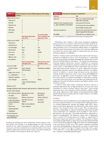
TABLE 33-1 Rapid Formulation of an Early Working Diagnosis of the Etiology TABLE 33-2 Indications for Intubation in Shock Patients
of Shock Indication Why
Defining features of shock
Hypoxemia High Fi O 2 is not guaranteed by oxygen
Blood pressure ↓ masks; PEEP can be added
Heart rate ↑ Ventilatory failure (inappropriately high Ensure adequate CO removal
2
Respiratory rate ↑ P CO 2 , signs of ventilatory muscle fatigue) Correct hypoxia due to hypoventilation
Mentation ↓ Prevent sudden respiratory arrest
Urine output ↓ Vital organ hypoperfusion Rest ventilatory muscles (and divert cardiac
Arterial pH ↓ output to hypoperfused vital organs)
Low Cardiac Output Obtundation Protect and ensure an adequate airway
High-Output Hypotension: Shock: Cardiogenic and FiO 2 , fraction of inspired O ; PCO 2 , partial pressure of CO2; PEEP, positive end-expiratory pressure.
Septic Shock Hypovolemic Shock 2
Is cardiac output reduced? No Yes
Pulse pressure ↑ ↓ Obtundation, due to shock or other causes, resulting in inadequate
airway protection is an important indication for intubation. In shock, air-
Diastolic pressure ↓ ↓ way intubation and mechanical ventilation should precede other compli-
Extremities/digits Warm Cool cated procedures, such as central venous catheterization, or complicated
tests that require transportation of the patient when these procedures and
Nail bed return Rapid Slow
tests restrict the medical staff’s ability to continuously assess the airway
Heart sounds Crisp Muffled and ensure adequacy of ventilation.
Temperature ↑ or ↓ ↔
Breathing: Initially, mechanical ventilation with sedation and, if necessary,
White cell count ↑ or ↓ ↔ paralysis are instituted to remove work of breathing as a confounding
Site of infection ++ — factor from the initial resuscitation and diagnostic pathway and to redis-
tribute limited blood flow to vital organs. The change from spontaneous
6
Reduced pump function, Reduced venous return, breathing (negative intrathoracic pressure ventilation) to mechanical
cardiogenic shock hypovolemic shock ventilation (positive intrathoracic pressure ventilation) leads to reduced
Is the heart too full? Yes No venous return so that additional volume resuscitation must be antici-
Symptoms, clinical Angina, abnormal Hemorrhage, dehydration pated when hypovolemia contributes to shock. Application of positive
context electrocardiogram end-expiratory pressure (increases intrathoracic pressure) and admin-
Jugular venous pressure ↑ ↓ istration of sedative or narcotic drugs (increases venous capacitance)
similarly should be expected to reduce venous return and highlight the
S , S , gallop rhythm +++ —
3 4 importance of aggressive volume resuscitation at the time of intuba-
Respiratory crepitations +++ — tion and institution of mechanical ventilation in hypovolemic patients.
Chest radiograph Large heart Normal Conversely, when hypovolemia is not a problem (eg, cardiogenic shock),
application of positive intrathoracic pressure may improve cardiac output
↑ Upper lobe flow Pulmonary and blood pressure.
edema
A relatively small tidal volume (6-8 mL/kg) should be selected to
What does not fit? minimize hypotension due to high intrathoracic pressures and, more
7
Overlapping etiologies (septic cardiogenic, septic hypovolemic, cardiogenic hypovolemic) importantly, to reduce ventilator-induced lung injury. When arterial
hypoxemia due to acute respiratory distress syndrome (ARDS) compli-
Short list of other etiologies
cates shock, adherence to tidal volumes of 6 mL/kg ideal body weight
High-output hypotension High right atrial pressure Nonresponsive significantly decreases mortality rate and number of days on a ventilator
Liver failure hypotension hypovolemia in the intensive care unit. 8
Severe pancreatitis Pulmonary hypertension Adrenal insufficiency Circulation
Trauma with significant (Most often pulmonary Anaphylaxis Goals and Monitoring Just as low tidal volumes limit ongoing lung inflammation
systemic inflammatory embolus) Spinal shock and injury, rapid resuscitation of the circulation limits ongoing generation
response Right ventricular infarction of a systemic inflammatory response and multiple organ injury. Hence,
Thyroid storm Cardiac tamponade rapid protocol-driven approaches with defined end points improve shock
2,3
Arteriovenous fistula outcome. For all types of shock, “time is tissue.” Thus, for hypovolemic
shock due to hemorrhage, the early goal is immediate hemostasis and rapid
Paget disease volume resuscitation. For cardiogenic shock secondary to acute myocar-
Get more information dial infarction, the early goal is immediate thrombolysis, angioplasty, or
9
Echocardiography, right surgical revascularization. For obstructive shock, relief of tamponade,
heart catheterization lysis or removal of the massive pulmonary embolus, or surgical relief of
abdominal compartment syndrome is required. The early goals of volume
resuscitation in hypovolemic or septic shock are incorporated in the Early
breathing precluding more than rudimentary verbal responses, tachy- Goal-Directed Therapy algorithm (Fig. 33-2), which was initially designed
pnea greater than 40/min or an inappropriately low and decreasing to aid resuscitation of septic shock. This requires immediate monitoring
3
respiratory rate, abdominal paradoxical respiratory motion, accessory (even before formal admission to the intensive care unit) of central venous
muscle use, and other manifestations of ventilatory failure such as inad- pressure (CVP; goal 8-2 mm Hg), MAP (goal >65 mm Hg), and Scv (goal
O 2
equately compensated acidemia should lead to early elective intubation >70%). When Scv is not readily measured then lactate clearance of >10%
O 2
and ventilation of the patient in shock (see Chap. 43). over ~2 hours is a reasonable alternative goal. 10
section03.indd 251 1/23/2015 2:06:53 PM