
Page 380 - Hall et al (2015) Principles of Critical Care-McGraw-Hill
P. 380
250 PART 3: Cardiovascular Disorders
is minimized by rapid and adequate (usefully driven by protocol) initial Interpretations of the data and response to initial therapy frequently con-
resuscitation. 2,3 firm the multiple etiologies or lead to a broader differential diagnosis of the
■ A QUESTIONING APPROACH TO THE INITIAL CLINICAL EXAMINATION etiologies of shock (see below). A short list of common etiologies other than
septic, hypovolemic, obstructive, or cardiogenic shock can be grouped as
Mean blood pressure is the product of cardiac output and systemic they present (see Table 33-1): high cardiac output hypotension that does
vascular resistance (SVR). Accordingly, hypotension may be caused by not appear to be caused by sepsis and poorly responsive hypovolemic shock.
reduced cardiac output or reduced SVR. Therefore, initial examination
of the hypotensive patient seeks to answer the question: Is cardiac output URGENT INITIAL RESUSCITATION
increased or decreased? (Fig. 33-1) High cardiac output hypotension is
most often signaled by a high pulse pressure, a low diastolic pressure, ■ PRIMARY SURVEY
warm extremities with good nail bed return, fever (or hypothermia), Early institution of aggressive resuscitation improves a patient’s chances
leukocytosis (or leukopenia), and other evidence of infection; these of survival. To improve efficiency at the necessarily rapid tempo, a
2,3
clinical findings strongly suggest a working diagnosis of septic shock systematic approach to initial evaluation and resuscitation is useful as
(Table 33-1), the initial treatment for which is thoughtful antibiosis it is during cardiac emergencies (advanced cardiac life support [ACLS])
combined with rapid expansion of the vascular volume and subsequent and trauma (advanced trauma life support [ATLS]). In analogy to these
vasopressors, inotropes, and blood transfusion as necessary to achieve an systematic “ABC” approaches, a primary survey includes establishing an
adequate central venous pressure (CVP), mean arterial pressure (MAP), airway (airway), choosing a ventilator mode and small tidal volumes that
; see below).
and central venous oxygen saturation (Scv O 2 minimize ventilator-induced lung injury (breathing), rapid (usefully pro-
In contrast, low cardiac output is signaled by a low pulse pressure, tocol driven) resuscitation of the inadequate circulation (circulation), and
mottled cyanotic skin, and cool extremities with poor nail bed return. drugs/definitive therapy consisting of early consideration and implemen-
In this case, clinical examination turns to a second question: Are central tation of definitive therapy for specific causes of shock (eg, hemostasis
veins empty or full? (Fig. 33-1) When low cardiac output results from for hemorrhage, revascularization for myocardial infarction, appropriate
hypovolemia (see Table 33-1) clinical examination shows manifestations antibiotics, surgical drainage of abscess, etc).
of blood loss (hematemesis, tarry stools, abdominal distention, reduced
hematocrit, or trauma) or manifestations of dehydration (reduced tis- Airway: Almost all patients in shock have one or more indications for air-
sue turgor, vomiting or diarrhea, or negative fluid balance). In contrast, way intubation and mechanical ventilation (Table 33-2), which should be
elevated jugular veins in a hypotensive patient suggest either obstruc- instituted early. Significant hypoxemia (based on blood-gas analysis, pulse
tion (eg, pulmonary embolism, cardiac tamponade) or cardiogenic oximetry, or high clinical suspicion) is one indication for airway intubation
shock (Fig. 33-1) raising the third question: Are breath sounds normal? because external masks and other devices do not reliably deliver an ade-
Cardiogenic shock is distinguished from obstructive shock by dependent quate fraction of inspired O (Fi O 2 ). Initially, a high Fi O 2 (100%) is used until
2
crackles on lung auscultation, a laterally displaced precordial apical blood-gas analysis or reliable pulse oximetry allows titration of the Fi O 2
impulse with extra heart sounds (S , S ), peripheral edema, chest pain, down toward less toxic concentrations.
3
4
ischemic changes on the electrocardiogram, and a chest radiograph show- Ventilatory failure is another indication for airway intubation and mecha-
ing a large heart with dilated upper lobe vessels and pulmonary edema. 4 nical ventilation. Elevated and rising partial pressure of CO in arterial
2
Whenever the clinical formulation is not obvious after answering the first blood reliably establishes the diagnosis of ventilatory failure but is often
three questions, ask a fourth: What does not fit? Most often, the answer is that a late finding. In particular, young, previously healthy patients are able
the hypotension is due to overlap of two or more of these common etiologies to defend partial pressure of CO (P CO 2 ) and pH up until a precipitous
2
of shock: septic shock complicated by hypovolemia or myocardial dysfunc- respiratory arrest. Therefore, clinical signs of respiratory muscle fatigue
tion, cardiogenic shock complicated by hypovolemia or sepsis, etc. At this or subtle evidence of inadequate ventilation are more important early
time, more data are frequently needed, especially aided by echocardiography. indicators. Evidence of respiratory muscle fatigue, including labored
5
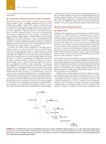
Increased Septic
Cardiac output? Empty Hypovolemic
Veins?
Decreased
Clear Obstructive
Full Breath sounds?
Dependent Cardiogenic
crackles
What does not fit?
Rare Kinds,
combinations
FIGURE 33-1. The different types of shock can be remembered using the SHOCK mnemonic and defined by asking four questions. First, is cardiac output increased (Septic shock) or
decreased (other forms)? Second, are central veins empty (Hypovolemic shock) or full (other forms)? Third, are breath sounds clear (Obstructive shock) or are crackles heard (Cardiogenic shock)?
Finally, what does not fit? This identifies combinations (eg, septic shock with hypovolemia) or rare kinds of shock. Additional physical findings, laboratory tests, and echocardiographic and other
examinations illuminate these simplified questions further.
section03.indd 250 1/23/2015 2:06:53 PM