
Page 400 - Hall et al (2015) Principles of Critical Care-McGraw-Hill
P. 400
270 PART 3: Cardiovascular Disorders
150 150
Doubled contractility Doubled contractility
Normal initial contractility
100 100 Low initial
LV Pressure LV Pressure contractility
50 50
0 0
0 50 100 150 0 50 100 150
12 12
10 10
Cardiac output 8 6 Doubled contractility Cardiac output 8 Doubled contractility
6
Normal initial contractility
4
4
Low initial
contractility
2 2
0 0
–5 0 5 10 15 20 25 –5 0 5 10 15 20 25
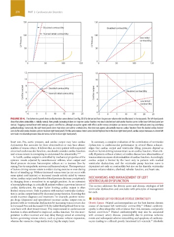
FIGURE 35-4. The bottom two panels show cardiac function curves derived (see Fig. 35-3 for derivation) from the pressure-volume relations illustrated in the top panels. The left-hand panels
show that when contractility is initially normal, then greatly increasing it does not improve cardiac function very much (dashed and solid cardiac function curves in the lower left-hand panel are
similar). Flogging a normal heart with inotropic agents is ineffective, although vasoactive agents with effects on the venous circulation can increase venous return without correcting underlying
pathophysiology. Conversely, the right-hand panels show that when contractility is initially low, then inotropic agents substantially improve cardiac function (from the dashed cardiac function
curve to the solid cardiac function curve in the lower right-hand panel). For the same venous return curve (dashed biphasic line in the lower right-hand panel), cardiac output increases at a lower left
ventricular end-diastolic pressure (blue dot versus red dot in lower right-hand panel).
heart rate, Pra, aortic pressure, and cardiac output may have cardiac In summary, a complete evaluation of the contribution of ventricular
dysfunction that accounts for these abnormalities or may have abnor- dysfunction to cardiovascular performance in critical illness acknowl-
malities of venous return. It follows that, in every patient with suspected edges that cardiac output and ventricular filling pressures depend as
abnormal cardiovascular function, one should consider cardiac function much on factors driving venous return as on cardiac function. Most criti-
and venous return in attempting to understand the abnormality. 21,23 cally ill patients without a history of cardiac disease have abnormalities of
In health, cardiac output is controlled by mechanical properties of the venous return in excess of abnormalities of cardiac function. Accordingly,
systemic vessels adjusted by neurohumoral reflexes; when output and cardiac output is limited by the heart only in patients with marked
blood pressure decrease, baroreceptor reflexes act to increase flow by ventricular dysfunction, and the ventricular pump function curve is
raising Pms by sympathetic nervous and humoral output. The importance dependent not only on contractility but also on the diastolic ventricular
of factors driving venous return is evident during exercise or even during pressure-volume relation, afterload, valvular function, and heart rate.
the act of standing up. Without increased venous tone (as can occur with
some spinal cord injuries) or increased muscle activity aided by venous
valves, cardiac output and therefore blood pressure decrease precipitously MECHANISMS AND MANAGEMENT OF LEFT
in changing from a recumbent to an upright position. As an extension VENTRICULAR DYSFUNCTION
of normal physiology, in critically ill patients without a previous history of This section addresses the diverse acute and chronic etiologies of left
cardiac dysfunction, the major factor limiting cardiac output is often ventricular dysfunction and concludes with principles of management
limited venous return. Only in patients with marked ventricular dysfunc- for each.
tion is cardiac output limited by decreased pump function. Knowing this
pic drugs (dopamine and epinephrine) increase cardiac output even in ■ DECREASED LEFT VENTRICULAR SYSTOLIC CONTRACTILITY
avoids incorrect diagnosis and treatment. For example, positive inotro-
patients with no ventricular dysfunction by increasing venous return due Chronic Causes: Dilated cardiomyopathies are the best-known chronic
to increased Pms and decreased RVR (by redistributing blood flow to vas- causes of decreased left ventricular contractility. Dilated cardiomy-
24
cular beds with short transit times). This improvement in cardiovascular opathy is often idiopathic with evidence that viral, immune, and genetic
function is often attributed to improved cardiac function. Yet this inter- factors contribute. Dilated cardiomyopathy may also be associated
24
pretation is often incorrect and may delay therapy aimed at correcting with coronary artery disease, presumably due to previous ischemic
factors governing venous return, such as plasma volume expansion, events and subsequent adverse remodeling and apoptosis of cardiomy-
25
whereas the vasoactive drugs ineffectively flog the empty heart. ocytes leading to a dilated, poorly functional left ventricle. Alcoholic
section03.indd 270 1/23/2015 2:07:03 PM