
Page 403 - Hall et al (2015) Principles of Critical Care-McGraw-Hill
P. 403
CHAPTER 35: Ventricular Dysfunction in Critical Illness 273
can also rapidly decrease ionized calcium levels and, as a result, may associated with an increase in lactic acid production because bicarbonate
depress ventricular contractility. In addition to ionized hypocalcemia, increases the rate-limiting step of glycolysis. Bicarbonate therapy also
42
other electrolyte abnormalities, including hypophosphatemia, hypo- decreases blood levels of ionized calcium. 41,42
magnesemia, and hypokalemia or hyperkalemia, may contribute to Decreased contractility due to ionized hypocalcemia can be corrected
decreased contractility or, more importantly, to arrhythmias. using an intravenous infusion of calcium. After approximately 6 U of
transfusion, ionized hypocalcemia should be measured and corrected, if
Management of Decreased Left Ventricular Contractility necessary. Hypophosphatemia, hypomagnesemia, hypokalemia, hyperka-
in Critical Illness lemia, and other metabolic disturbances should also be corrected because
Identify and Correct Acute Reversible Causes It is important to identify the mul- they may lead directly or indirectly to altered cardiovascular function.
tiple, different potentially reversible causes for depressed contractility in Managing the Depressed Heart Having reversed the acute contributors to
critically ill patients because, although alone they may be insufficient to depressed left ventricular contractility, standard therapy of decreased left
account for the left ventricular dysfunction, together they may signifi- ventricular contractility includes optimizing ventricular filling pressure,
cantly depress function. For example, if ischemia or hypoxemia is pres- decreasing afterload when arterial pressure is adequate, and increasing
ent, aggressive attempts to correct it should be instituted. In the presence contractility using inotropic agents. 45,46 These therapies are considered
of coronary artery disease, standard care including heparin, antiplatelet in detail below (see “Acute on Chronic Heart Failure”). Assisted positive
therapy, β-blockade, and coronary vasodilation using nitrates may be pressure ventilation may help by improving oxygenation, decreasing
helpful. Emergency percutaneous coronary intervention (PCI) to rapidly dyspnea, and decreasing left ventricular afterload. This can be applied
reestablish perfusion or, if this is not possible, early thrombolytic therapy using noninvasive mask ventilation (eg, CPAP or BiPAP) or, following
after acute coronary thrombosis decreases the incidence of congestive intubation, using conventional mechanical ventilation modes. 47,48
heart failure and improves outcome (see Chap. 37). When vasodilator and inotropic therapy is insufficient, temporary
Limitation of the intramyocardial inflammatory response by early support using intra-aortic balloon counterpulsation, ventricular assist
aggressive treatment of myocardial ischemia or by early aggressive treat- devices, or ECMO is appropriate when damaged myocardium is
ment of sepsis and its associated systemic inflammatory response are expected to recover or as supportive therapy leading to surgical correc-
effective. 43,44 Specific treatment using anti-inflammatory strategies has tion of an anatomic abnormality (see Chap. 53). 49,50 Resynchronization
not yet been shown to definitively improve ventricular function. therapy using biventricular pacing may help. Finally, heart transplan-
51
Correction of hypoxemia and anemia may result in substantial tation may be necessary. Stem cell transplantation is a promising new
improvement in ventricular function. Attention should be paid to approach under investigation to repair damaged myocardium. 52,53
decreasing factors that increase myocardial oxygen demand. Therefore, The ventricular pump function curve illustrates the Frank-Starling
when β-blockade is not feasible, choosing the lowest level of inotropic mechanism, which shows that increased ventricular filling results
and vasoactive drugs that produces the desired therapeutic effect will in increased ejection even when contractility is depressed. The limit to
minimize their contribution to myocardial oxygen demand. Likewise, increased ventricular filling is generally set by the onset of pulmonary
alleviating pain is important to diminish the associated tachycardia and edema. Pulmonary edema fluid enters the lung interstitium according to
increased sympathetic tone. the Starling equation. At normal protein osmotic pressures (largely due
In ventilated patients with left ventricular dysfunction, the detrimental to albumin) and normal permeability of the pulmonary endothelium, pul-
effects of acute respiratory acidosis should be considered; mixed venous monary edema starts to develop at Ppw values of at least 20 to 25 mm Hg.
54
and, hence, tissue P is much higher than the arterial partial pressure of In the presence of decreased oncotic pressure due to decreased albumin
CO 2
CO when the cardiac output is low. In general, metabolic acidosis should or in the presence of a leaky pulmonary endothelium, pulmonary edema
2
be treated by reversing its etiology. Alkali therapy for increased anion gap may form at considerably lower Ppw values; in acute respiratory distress
metabolic acidosis is of no benefit and may be dangerous even at pH values syndrome (ARDS) or pneumonia, pulmonary edema may form at very
as low as 7.0 for a number of reasons. Bicarbonate infusion results in low Ppw values. With this in mind, it is appropriate to search for the Ppw
42
an increase in P due to chemical equilibrium of HCO with H O and that produces the highest cardiac output without resulting in substantial
−
3
2
CO 2
CO unless compensatory hyperventilation is also instituted. Particularly pulmonary edema. Most often, this search necessitates preload reduction
2
during rapid bolus injection, local P may climb to extremely high using diuretics and vasodilating agents (Table 35-3). However, when pul-
CO 2
values so that myocardial intracellular acidosis transiently may be severe, monary edema does not limit oxygenation, it is appropriate to consider
leading to decreased ventricular contractility. Bicarbonate therapy is increasing cardiac output by intravascular fluid expansion.
37
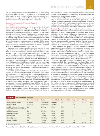
TABLE 35-3 Effect of Direct-Acting Vasodilators
Drug Route of Administration Dosage Onset of Effect Duration of Effect Large Arteries Arterioles Veins
Sodium nitroprusside (Nipride) Intravenous 25-400 µg/min Immediate — + +++ +++
Nitroglycerin (Tridil) Intravenous 10-200 µg/min Immediate — ++ + +++
Isosorbide dinitrate (Isordil; Sorbitrate, Oral 20-60 mg 30 min 4-6 h ++ + +++
Isobid; Isotrate; Sorate, Sorbide; Dilatrate)
Hydralazine (Apresoline) Oral 50-100 mg 30 min 6-12 h 0 +++ ±
Hydralazine (Apresoline) IV or IM 5-40 mg 15 min 4-8 h 0 +++ ±
Minoxidil (Loniten) Oral 10-30 mg 30 min 8-12 h 0 +++ 0
Diazoxide (Hyperstat) IV bolus 100-300 mg Immediate 4-12 h 0 +++ ±
Nifedipine (Procardia) Oral 10-20 mg 20-30 min 2-4 h ++ +++ ±
Sublingual 10-20 mg 15 min 2-4 h ++ +++ ±
IM, intramuscular; IV, intravenous.
Reproduced with permission from Cohn JN. Drugs used to control vascular resistance and capacitance. In: Hurst JW, et al, eds. The Heart, Arteries and Veins. 7th ed. New York: McGraw-Hill; 1990.
section03.indd 273 1/23/2015 2:07:05 PM