
Page 397 - Hall et al (2015) Principles of Critical Care-McGraw-Hill
P. 397
CHAPTER 35: Ventricular Dysfunction in Critical Illness 267
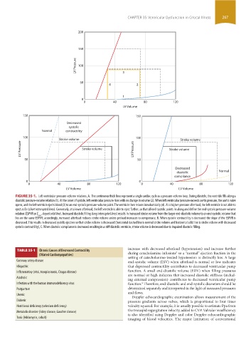
200
150
LV Pressure 100 3
50
4 2
1
0
0 40 80 120
LV Volume
150 150
Decreased
systolic
Normal contractility
100 Stroke volume 100 Stroke volume
LV Pressure Stroke volume LV Pressure Stroke volume
50 50
Decreased
diastolic Normal
compliance
0 0
0 40 80 120 0 40 80 120
LV Volume LV Volume
FIGURE 35-1. Left ventricular pressure-volume relations, A. The continuous thick lines represent a single cardiac cycle as a pressure-volume loop. During diastole, the ventricle fills along a
diastolic pressure-volume relation (1). At the onset of systole, left ventricular pressure rises with no change in volume (2). When left ventricular pressure exceeds aortic pressure, the aortic valve
opens, and the left ventricle ejects blood (3) to an end-systolic pressure-volume point. The ventricle then relaxes isovolumically (4). At a higher-pressure afterload, the left ventricle is not able to
eject as far (short interrupted lines). Conversely, at a lower afterload, the left ventricle is able to eject farther, so that all end-systolic points lie along and define the end-systolic pressure-volume
relation (ESPVR or E , sloped solid line). Increased diastolic filling (long interrupted lines) results in increased stroke volume from the larger end-diastolic volume to an end-systolic volume that
max
lies on the same ESPVR; accordingly, increased afterload reduces stroke volume unless preload increases to compensate, B. When systolic contractility is decreased the slope of the ESPVR is
decreased. This results in decreased systolic ejection so that stroke volume is decreased (horizontal dashed line is normal stroke volume and horizontal solid line is stroke volume with decreased
systolic contractility), C. When diastolic compliance is decreased resulting in a stiff diastolic ventricle, stroke volume is decreased due to impaired diastolic filling.
increase with decreased afterload (hypotension) and increase further
TABLE 35-1 Chronic Causes of Decreased Contractility during catecholamine infusions so a “normal” ejection fraction in the
7
(Dilated Cardiomyopathies)
setting of catecholamine-treated hypotension is distinctly low. A large
Coronary artery disease end-systolic volume (ESV) when afterload is normal or low indicates
Idiopathic that depressed contractility contributes to decreased ventricular pump
Inflammatory (viral, toxoplasmosis, Chagas disease) function. A small end-diastolic volume (EDV) when filling pressures
are normal or high indicates that increased diastolic stiffness (includ-
Alcoholic
ing external compression) contributes to decreased ventricular pump
Infection with the human immunodeficiency virus function. Therefore, end-diastolic and end-systolic diameters should be
8
Postpartum determined separately and interpreted in the light of measured pressures
Uremic and flows.
Doppler echocardiographic examination allows measurement of the
Diabetic pressure gradients across valves, which is proportional to four times
Nutritional deficiency (selenium deficiency) velocity squared. For example, it is usually possible to estimate Ppa from
Metabolic disorder (Fabry disease, Gaucher disease) the tricuspid regurgitation velocity, added to CVP. Valvular insufficiency
is also identified using Doppler and color Doppler echocardiographic
Toxic (Adriamycin, cobalt) imaging of blood velocities. The major limitation of conventional
section03.indd 267 1/23/2015 2:07:00 PM