
Page 442 - Hall et al (2015) Principles of Critical Care-McGraw-Hill
P. 442
312 PART 3: Cardiovascular Disorders
accurate. However in a meta-analysis of 23 studies that included
TABLE 38-3 Causes of RV Failure in ICU
807 patients, 3D TTE underestimated RV volumes and EF when com-
RV Pressure Overload, Pulmonary Hypertension, Any Cause pared with the gold-standard measurements by cardiac MRI. 43
Pulmonary embolism
Pulmonary Artery Catheterization: Pulmonary artery catheterization
ARDS can estimate pulmonary arterial pressures more accurately than echo-
Excessive PEEP, tidal volume, and alveolar pressure cardiography. However, interpretation of mean pulmonary pressures
and measurement of tricuspid regurgitation by thermodilution are
Air, amniotic, fat, or tumor microembolism
confounded by technical limitations. RV failure is characterized by a
Sepsis (rarely) reduced cardiac output (typically cardiac index <2.5 L/min/m ) and
2
Pulmonary leukostasis, leukoagglutination an elevation in right sided filling pressures (eg, right atrial pressure
Extensive lung resection >8 mm Hg). A pulmonary artery catheter (PAC) with a fast-response
thermistor has been advocated for accurate measurement of right ven-
Drugs (eg, heparin-protamine reaction) tricular end-diastolic volume (RVEDV) and hemodynamic parameters
Hypoxia including RV ejection fraction by thermodilution in the presence of
Reduced RV Contractility tricuspid regurgitation. However the fast-response thermistor PAC may
systematically overestimate RVEDV in the presence of ischemia and
44
RV infarction
has not been demonstrated to confer an improvement in survival.
Sepsis
Circulating Biomarkers: The utility of cardiac biomarkers for diag-
RV cardiomyopathy
nosing acute RV injury in RHS has been demonstrated (mainly in
Myocarditis pericardial disease; LVAD; post-CPB; postcardiac surgery/transplantation acute PE) to accurately identify low-risk patients. BNP assay nega-
RV-Volume Overload tive predictive values for in-hospital death range from 97% to 100%.
RV systolic failure is an independent determinant of serum levels of
Tricuspid and pulmonary regurgitation; intracardiac shunts
brain natriuretic peptide (BNP) in patients with severe heart failure.
35
ARDS, acute respiratory distress syndrome; HIV, human immunodeficiency virus; Pa, pulmonary artery; However, the performance characteristics (positive predictive value
PEEP, positive end-expiratory pressure; RV, right ventricular. and sensitivity) are inconsistent and preclude the use of either BNP
Data from Price LC, Wort SJ, Finney SJ, et al. Pulmonary vascular and right ventricular dysfunction in adult critical or BNP levels across as range of cutoff values for routine diagnosis or
care: current and emerging options for management: a systematic literature review. Crit Care. 2010;14(5):R169. prognosis in patients with moderate to high pretest probability. 5
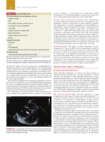
the typical “D” shape of the LV on the short-axis view (Fig. 38-4); para- SPECIFIC RIGHT HEART SYNDROMES
doxical septal motion in systole; right Pa dilation; or loss of respirophasic
variation in the inferior vena cava. RV infarction can usually be readily ■ ACUTE PULMONARY HYPERTENSION
39
distinguished from acute pulmonary hypertension in that high Pa pres-
sures are lacking. Right ventricular diastolic dimensions can be obtained Acute pulmonary hypertension is caused by an abrupt increase in
by measuring right ventricular end-diastolic area in the long axis, pulmonary vascular resistance due to vascular obstruction or surgical
from an apical four-chamber view, or by a transesophageal approach in resection. The prototype of acute pulmonary hypertension is acute
the volume-repleted patient. 40 pulmonary embolism (PE; see Chap. 39), but other forms of embolism
Enhanced echo techniques that are independent of geometrical assump- (eg, air or fat), microvascular injury (eg, ARDS), drug effect, and inflam-
tions have been developed to assess acute pathophysiological changes in mation can acutely raise pulmonary vascular resistance (see Table 38-3).
RV function. Tricuspid annular plane systolic excursion (TAPSE), RV In its most severe form, acute pulmonary hypertension associated with
41
systolic and diastolic tissue Doppler imaging (TDI) velocities and Speckle profound RV dysfunction is termed acute cor pulmonale. 11,45,46 The echo-
tracking-derived strain TAPSE has been demonstrated to be a sensitive cardiographic diagnosis of acute cor pulmonale consists of the combina-
42
marker of acute RV dysfunction in 40 patients with acute PE. 41 tion of RV dilation (reflecting RV diastolic overload) with paradoxical
Newer algorithms for assessment of RV volumes and ejection frac- septal motion during systole (reflecting RV systolic overload). 32
tion by real-time three-dimensional TTE are reported to be reasonably
Right Ventricular Infarction: Right ventricular infarction is a well-
recognized and fatal feature of inferior myocardial infarction. 47,48 It
is also seen in anterior infarcts. In most cases RV free wall infarction
or ischemia is accompanied by varying degrees of septal and pos-
teroinferior left ventricular injury, but relatively isolated RV injury is
occasionally seen. RV myocardial injury and dysfunction represent-
ing noninfarcted hibernating myocardium may be able to sustain
long periods of low coronary oxygen delivery and ultimately recover
substantial contractile function. 49
RV dilation accompanies significant myocardial injury. Concomitant
LV infarction involving the interventricular septum may lead to further
hemodynamic deterioration in patients with RV infarction because of
the loss of LV septal contraction, which can assist RV ejection. Elevation
of right atrial pressure on physical examination or direct measurement
in a patient with an inferior myocardial infarction and clear lungs by
exam and chest x-ray should lead to suspicion of RV infarction. When
these features occur in a critically ill patient, the essential distinction is
between RHS resulting from acute Pa hypertension and RHS resulting
from RV infarction. Confirmatory evidence includes a right precordial
FIGURE 38-4. Echocardiographic short-axis view showing the obvious shift of the inter- electrocardiogram or echocardiographic evidence of RV injury (see
ventricular septum toward the left ventricle, changing the shape of the left ventricle from its Chap. 37). Proximal RCA occlusion commonly results in concomitant
normal circular cross-section to a “D” shape. right atrial ischemia. This can precipitate significant rate and rhythm
section03.indd 312 1/23/2015 2:07:27 PM