
Page 440 - Hall et al (2015) Principles of Critical Care-McGraw-Hill
P. 440
310 PART 3: Cardiovascular Disorders
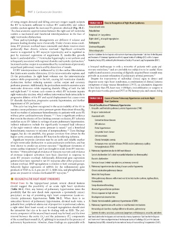
of rising oxygen demand and falling coronary oxygen supply subjects TABLE 38-1 Clues to Recognition of Right Heart Syndromes
the RV to ischemia sufficient to reduce RV contractility and reduce
systolic ejection against the increased Pa pressure afterload (Fig. 38-1). Elevated neck veins
The close anatomic approximation between the right and left ventricles Pulsatile liver
confers a mechanical and functional interdependence in the face of Peripheral ≫ lung edema
right ventricular dysfunction.
These pathophysiologic derangements are different if volume and Right sided S , tricuspid regurgitation
3
pressure loading develop more chronically. Under chronic stress condi- Radiographic
tions, RV pressure overload taxes contractile and elastic reserves more Electrocardiographic
profoundly than chronic volume overload. Significant contractile
7
reserve is supported by RV myocyte hypertrophy and is regulated in Echocardiographic
part by increased expression of angiotensin II, insulin-like growth Data from Guidelines for the diagnosis and treatment of pulmonary hypertension: Task Force for the Diagnosis
factor-I, and endothelin-1. Ventricular hypertrophy is not uniform and and Treatment of Pulmonary Hypertension of the European Society of Cardiology (ESC) and the European
8
is frequently associated with regional diastolic and systolic dysfunction. Respiratory Society (ERS), endorsed by the International Society of Heart and Lung Transplantation (ISHLT).
9
Increased cardiac output is accommodated by recruitment of previously
unperfused pulmonary vessels and by distention of vessels. a binaural stethoscope in only a minority of patients with acute pul-
18
LV/RV interdependence is influenced by (1) the cardiac fibroskeleton monary embolism, and are probably too subjective to be useful. More
that limits acute annular distension, (2) the interventricular septum, and sophisticated acoustic processing of digitally acquired heart sounds may
(3) the pericardium. As right heart volumes rise, the interventricular provide an accurate estimation of pulmonary arterial pressures. 19
septum shifts progressively to the left, causing left ventricular diastolic Despite the insensitivity of individual clinical signs to detect and
dysfunction, further reducing systemic cardiac output and coronary diagnose acute right heart syndromes, a combination of clinical features
perfusion pressure. Additionally, the pericardium restricts excessive acute (symptoms of deep venous thrombosis [DVT]; an alternative diagnosis
ventricular distension while impairing diastolic filling of both the left is less likely than PE; heart rate >100 bpm; immobilization or surgery in
and right heart. A vicious cycle ensues in which RV ischemia impairs the previous 4 weeks; previous DVT or PE; hemoptysis; and cancer, being
10
right ventricular ejection, which without intervention leads intractably to
progressive dilation of the RV and septal displacement that causes more TABLE 38-2 Causes of Severe Pulmonary Hypertension and Acute Right
LV diastolic dysfunction, progressive systemic hypotension, and further Heart Syndrome
impairment of RV perfusion. 11
This cycle has long been recognized in the acute inability of the RV to Clinical Classification of Pulmonary Hypertension
sustain a mean pulmonary artery pressure greater than about 40 mm Hg, 1. Idiopathic and heritable pulmonary arterial hypertension (PAH)
based on studies of pulmonary hemodynamics in patients with acute PE Drugs or toxins induced
without prior cardiopulmonary disease. There is significant evidence Associated with:
1,12
that even in the absence of flow limiting coronary occlusion, RV ischemia
underlies acute RV failure in settings of acute pulmonary hypertension. Connective tissue diseases
Indirect indications include the significantly increased load tolerance HIV infection
of the right ventricle when aortic pressure is raised, and a beneficial Portal hypertension
13
hemodynamic response to infusion of norepinephrine. These findings
14
suggest, but do not establish, that greater coronary flow driven by the Congenital heart disease
higher aortic pressure enhances RV function by relieving ischemia. Schistosomiasis
Significant troponin elevation may be an early and reliable marker Chronic hemolytic anemia
of right ventricular dysfunction in acute pulmonary embolism, and has Pulmonary veno-occlusive disease (PVOD) and/or pulmonary capillary
been shown to predict an adverse outcome. Significant elevations of hemangiomatosis (PCH)
15
serum cardiac troponins T and I are thought to result from RV microin-
farction. Histopathological evidence of myocyte necrosis and evidence 2. Pulmonary hypertension due to left heart disease
16
of protease (calpain) activation have been described in response to Systolic dysfunction (left-sided myocardial infarction/cardiomyopathy)
acute RV pressure overload. Additionally differential gene expression Diastolic dysfunction
patterns have been reported in rat RV myocytes after either pressure or
volume overload. BNP upregulation is evident in both stressed groups. Valvular disease (mitral regurgitation; pulmonary stenosis)
Relatively higher expression of mRNA for the inflammatory gene 3. Pulmonary hypertension due to lung diseases and/or hypoxemia
products of TNF-α, IL-6, pre-pro ET-1, SERCA2a, and phospholamban Chronic obstructive pulmonary disease
genes are present in volume overloaded RV myocytes. 17 Interstitial lung disease
■ RECOGNIZING THE RIGHT HEART SYNDROMES Other pulmonary diseases with mixed restrictive/obstructive pattern, kyphoscoliosis,
Clinical Clues: In the hypoperfused patient, several clinical features thoracoplasty
should suggest the possibility of an acute right heart syndrome Sleep-disordered breathing
(Table 38-1). First, any history of pulmonary hypertension raises the Alveolar hypoventilation syndrome
possibility that the new shock state represents a (potentially minor) Chronic exposure to high altitude
precipitant on top of preexisting right heart compromise (acute-on-
chronic pulmonary hypertension; Table 38-2). When there is no Developmental abnormalities
antecedent history of pulmonary hypertension, elevated neck veins, a 4. Chronic thromboembolic pulmonary hypertension (CTEPH)
pulsatile liver, peripheral edema out of proportion to pulmonary edema, 5. Pulmonary hypertension with unclear or multifactorial mechanisms
a right-sided third heart sound, or tricuspid regurgitation should alert
the intensivist that she or he may be dealing with an RHS. The pul- Hematological disorders: myeloproliferative disorders, splenectomy
monic component of the second heart sound may be loud, and the time Systemic disorders, sarcoidosis, pulmonary Langerhans cell histiocytosis, vasculitis, and others
interval between the aortic (A ) and the pulmonary (P ) components Data from Guidelines for the diagnosis and treatment of pulmonary hypertension: Task Force for the Diagnosis
2
2
of the second heart sound (A -P splitting) is increased in the presence of and Treatment of Pulmonary Hypertension of the European Society of Cardiology (ESC) and the European
2
2
pulmonary hypertension. However, these findings are appreciable with Respiratory Society (ERS), endorsed by the International Society of Heart and Lung Transplantation (ISHLT).
section03.indd 310 1/23/2015 2:07:25 PM