
Page 456 - Hall et al (2015) Principles of Critical Care-McGraw-Hill
P. 456
326 PART 3: Cardiovascular Disorders
■
or indeterminate results. Scans of intermediate probability indicate EMPIRICAL DIAGNOSIS
a substantial likelihood of PE (~40%) necessitating further evalua- Occasionally, an empirical diagnosis of PE seems clear cut to the manag-
tion to prove or exclude the diagnosis. Furthermore, a scan read as ing physician. No alternative diagnoses may seem plausible, or further
“low probability” is not helpful in critically ill patients, since this diagnostic steps seem risky or unnecessary. Although this approach may
group rarely has a low pretest probability for PE, and PE prevalence appear attractive, it has attendant drawbacks. In the critically ill popula-
in this group can again approach 40%. In practice, it would seem tion, there will remain competing alternative diagnoses, and the clinical
that the utility of V/Q scanning is largely limited to patients with an diagnosis remains difficult, even for the more experienced clinician.
imperative either to avoid intravenous contrast due to allergy or renal Most important, the doubt which lingers after an empirical diagnosis too
impairment, or to minimize radiation, such as in the case of preg- frequently haunts subsequent management. Progression of symptoms
nancy, which is discussed below. Furthermore, patients who have a or signs despite therapy raises questions about failure of treatment or
normal chest are the most likely to have an interpretable scan.
https://kat.cr/user/tahir99/
the need for alternative treatments. Complications of treatment such as
Other Noninvasive Means of Diagnosis: In noncritically ill patients, the hemorrhage or thrombocytopenia create uncertainty about the neces-
D-dimer assay has proven to be a very helpful noninvasive test. D-dimer sity of the toxic therapy, or precipitate more diagnostic interventions in
is a fibrin degradation product that appears in the blood when there is a newly unstable state. Since critically ill patients are more likely to have
some degree of fibrinolysis. Very low levels of D-dimer argue against complications of therapy, long-term empiricism is rarely appropriate.
a diagnosis of PE, with a sensitivity of 95% and a negative likelihood Instead, initial empiricism while the patient is stabilized should give
ratio of approximately 0.10 using the widely available quantitative rapid way to appropriate diagnostic testing as the patient’s condition improves.
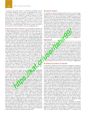
ELISA. Essential to the use of a D-dimer test in diagnosing patients with A diagnostic algorithm tailored to the ICU is presented in Figure 39-5.
57
thromboembolic disease is an assessment of clinical pretest probability;
for patients assessed as intermediate or high risk based on clinical factors, TREATMENT
a negative D-dimer could still result in up to 25% of the patients having The majority of patients with PE will not die from the clot which
a PE. The test is thus advocated only for clinically low-risk patients, and leads to diagnosis. As long as reembolization is prevented, the patient
some experts caution against using it at all for hospitalized patients. will survive, while intrinsic fibrinolysis restores pulmonary blood
44
Furthermore, D-dimer assays can be considered unidirectional, in that a flow. Therefore, the primary goal of all therapies for PE is to prevent
negative result can be extremely useful, yet a positive result has little effect reembolization. Some patients, however, survive the initial embolus,
on the likelihood of either DVT or PE. In the critically ill population, yet remain in shock. These patients, who are overrepresented in ICU
57
few patients would be characterized as low risk for PE and almost all populations, may succumb to the initial embolus. Additional therapy to
58
patients will have a positive D-dimer level, rendering the test unhelpful. hasten clot resolution, aimed at more promptly restoring the circulation,
59
The combination of pulse oximetry and static compliance of the in addition to supportive care for the strained right ventricle, is useful in
respiratory system yielded a very high sensitivity and specificity for PE such patients. Beyond anticoagulation, vena caval interruption, throm-
in critically ill trauma patients. In patients with COPD, capnography bolysis, fluid and vasoactive drug administration, and rarely, surgical
60
and arterial blood gas demonstrating a low dead space fraction had embolectomy all may be considered in the treatment of this disease. An
a very strong negative predictive value for PE. The combination of integrated approach to the treatment of PE is presented in Figure 39-6.
15
16 ■
steady-state end-tidal alveolar dead space fraction and D-dimer was also
quite sensitive in diagnosing PE in hospitalized, though not critically PROGNOSIS AND INTENSITY OF TREATMENT
ill, patients, with a sensitivity and negative predictive value of 98%.
Whether these tests can be applied in critically ill patients with addi- Having made a diagnosis of PE, the clinician and patient face numerous
tional cardiopulmonary derangements remains less certain. potential therapies and outcomes. Pulmonary embolism is spectacularly
inconsistent in its clinical presentation, and can range from asymptomatic
Pulmonary Angiography: Digital subtraction pulmonary angiography or mildly symptomatic dyspnea to profound shock due to right ventricu-
(DSA) has long been considered the definitive test for the diagnosis of lar dysfunction. Several characteristics of each presentation can allow the
PE. Positive findings include an intraluminal filling defect or a cutoff of a clinician to identify patients with the poorest prognosis, who almost cer-
2-mm or larger vessel seen in more than one view. Experienced radiologists tainly benefit from close observation in a monitored setting, and who may
61
agree on 98% of studies showing lobar embolism. However, agreement benefit from a more aggressive therapeutic approach. Equally important,
falls to 90% with segmental embolism, and only 66% in those with the clinician may also identify those patients at low risk for complication, in
subsegmental clots, again highlighting the diagnostic challenge of small whom a strategy of anticoagulation alone, potentially as an outpatient, will
pulmonary thrombi, and uncertainty surrounding their clinical significance. suffice. The Geneva prognostic index, generated from a prospective study
Following a negative DSA, the risk of subsequent VTE is less than 2%. 62-64 of 296 patients with PE admitted through the emergency room, identified
Because the earliest documented resolution of an angiogram to normal six predictors of adverse outcome, defined as death, recurrent thrombotic
following a pulmonary embolus is 1 week, there may not be time urgency event, or major bleeding. Hypotension imparted an odds ratio of 15 for
69
in performing the test as long as anticoagulation can be empirically started, adverse event; cancer of 9.5; and prior DVT, DVT by ultrasound, heart
and the result of DSA appears to be reliable up to a week following acute failure, and hypoxemia increased odds in the range of two- to fourfold.
symptoms. Interestingly, in a retrospective review of the 20 discordant More recently, the simplified PE severity index (sPESI) was shown to
65
cases between CTPA and DSA in the PIOPED II study, it was determined identify a subgroup of PE patients with a low 30-day mortality (1%) in
that CTPA had a superior sensitivity, with 2 false-negatives compared both a discovery and large validation cohort. Low risk patients were
70
to 13 for DSA. Since it is invasive, costly, riskier, and involves more those with none of the following criteria: age >80 years; history of cancer;
66
radiation than CT angiography, DSA is usually reserved for patients in history of chronic cardiopulmonary disease; heart rate ≥110 beats/min;
61
whom the diagnosis cannot be made or excluded by less invasive means. systolic blood pressure <100 mm Hg; or arterial O saturation <90%.
2
However, pulmonary angiography appears to be safer than is generally Some have advocated using this low risk group to determine which
appreciated. In several large series, mortality was approximately 0.2%. 67,68 patients can be safely treated with LMWH as an outpatient. 71
Case reports of death periangiography often cite pulmonary hypertension In the ICU, the more common scenario is attempting to identify
and cor pulmonale at the time of the procedure, leading some to conclude patients at high risk for adverse events, in order to provide more intensive
that severe pulmonary hypertension is a contraindication to pulmonary monitoring and to prepare for escalation of therapy if necessary. Plasma
angiography. Elevated pulmonary systolic pressure (>70 mm Hg) and markers of cardiac injury such as troponin T and troponin I portend a
elevated right ventricular diastolic pressure (>20 mm Hg) were identified high risk for complications, and troponin I was significantly associated
as risk factors for death, with a reported mortality of 2%. 68 with an increased overall mortality following PE. For patients with
72
section03.indd 326 1/23/2015 2:07:35 PM