
Page 454 - Hall et al (2015) Principles of Critical Care-McGraw-Hill
P. 454
324 PART 3: Cardiovascular Disorders
high degree of reliability. While CTPA protocol is minimally invasive, it
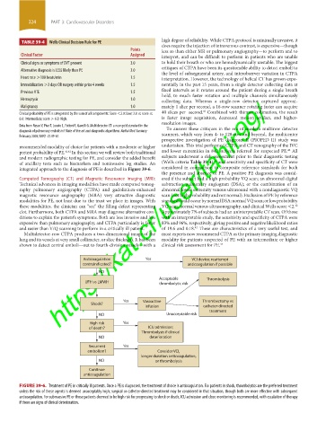
TABLE 39-4 Wells Clinical Decision Rule for PE
does require the injection of intravenous contrast, is expensive—though
Points less so than either MR or pulmonary angiography—to perform and to
Clinical Factor Assigned interpret, and can be difficult to perform in patients who are unable
Clinical signs or symptoms of DVT present 3.0 to hold their breath or who are hemodynamically unstable. The biggest
critiques of CTPA have been its questionable ability to detect emboli to
Alternative diagnosis is LESS likely than PE 3.0
the level of subsegmental artery, and interobserver variation in CTPA
Heart rate >100 beats/min 1.5 interpretation. However, the technology of helical CT has grown expo-
Immobilization >3 days OR surgery within prior 4 weeks 1.5 nentially in the past 15 years, from a single detector collecting data at
fixed intervals as it rotates around the patient during a single breath
Previous VTE 1.5
hold, to much faster rotation and multiple channels simultaneously
https://kat.cr/user/tahir99/
Hemoptysis 1.0 collecting data. Whereas a single-row detector captured approxi-
Malignancy 1.0 mately 1 slice per second, a 16-row scanner rotating faster can acquire
43
Clinical probability of PE is categorized by the sum of all components: Score <2.0: Low; 2.0 ≤ score ≤ 40 slices per second. Combined with thinner collimation, the result
6.0: Intermediate; score > 6.0: High. is faster image acquisition, decreased motion artifact, and higher-
resolution images.
Data from Russo V, Piva T, Lovato L, Fattori R, Gavelli G. Multidetector CT: a new gold standard in the To answer these critiques in the era of modern multirow detector
diagnosis of pulmonary embolism? State of the art and diagnostic algorithms. Radiol Med. January- scanners, which vary from 4- to 128-row and beyond, the multicenter
February 2005;109(1-2):49-61.
prospective investigation of PE diagnosis II (PIOPED II) study was
recommended modality of choice for patients with a moderate or higher undertaken. This trial performed CTPA and CT venography of the IVC
42
pretest probability of PE. 41,42 In this section we will review both traditional and lower extremities in 842 subjects referred for suspected PE. All
and modern radiographic testing for PE, and consider the added benefit subjects underwent a risk assessment prior to their diagnostic testing
40
of ancillary tests such as biomarkers and noninvasive leg studies. An (Wells criteria Table 39-4), and sensitivity and specificity of CT were
integrated approach to the diagnosis of PE is described in Figure 39-6. considered in comparison to composite reference standards for both
the presence and absence of PE. A positive PE diagnosis was consid-
Computed Tomography (CT) and Magnetic Resonance Imaging (MRI): ered if the subject had a high probability VQ scan; an abnormal digital
Technical advances in imaging modalities have made computed tomog- subtraction pulmonary angiogram (DSA); or the combination of an
raphy pulmonary angiography (CTPA) and gadolinium-enhanced abnormal lower extremity venous ultrasound with a nondiagnostic VQ
magnetic resonance angiography (MRA) very attractive diagnostic scan (not high probability and not normal). Exclusion of PE by reference
modalities for PE, not least due to the trust we place in images. With standard could occur by normal DSA; normal VQ scan; or low probability
these modalities, the clinician can “see” the filling defect representing VQ scan, normal venous ultrasonography, and clinical Wells score <2.
42
clot. Furthermore, both CTPA and MRA may diagnose alternative con- Approximately 7% of subjects had an uninterpretable CT scan. Of those
ditions to explain the patient’s symptoms. Both are less invasive and less with an interpretable study, the sensitivity and specificity of CTPA were
expensive than pulmonary angiography, and CTPA particularly is faster 83% and 96%, respectively, giving positive and negative likelihood ratios
and easier than V/Q scanning to perform in a critically ill patient. of 19.6 and 0.18. These are characteristics of a very useful test, and
42
Multidetector-row CTPA produces a two-dimensional image of the most experts now recommend CTPA as the primary imaging diagnostic
lung and its vessels at very small collimeter, or slice thickness. It has been modality for patients suspected of PE with an intermediate or higher
shown to detect central emboli—out to fourth-division vessels—with a clinical risk assessment for PE. 44
Anticoagulation Yes VCI device; reattempt
contraindicated? anticoagulation if possible
NO
Acceptable Thrombolysis
UFH vs LMWH thrombolytic risk
Yes Vasoactive Thrombectomy vs
Shock?
infusion catheter-directed
treatment
NO Unacceptable risk
High risk Yes
of death? ICU admission;
Thrombolysis if clinical
NO deterioration
Recurrent Yes
embolism? Consider VCI,
longer duration anticoagulation,
NO or thrombolysis
Continue
anticoagulation
FIGURE 39-6. Treatment of PE in critically ill patients. Once a PE is diagnosed, the treatment of choice is anticoagulation. For patients in shock, thrombolytics are the preferred treatment
unless the risk of these agents is deemed unacceptably high; surgical or catheter-directed treatment may be considered in that situation, though both are more effective with subsequent
anticoagulation. For submassive PE or those patients deemed to be high risk for progressing to shock or death, ICU admission and close monitoring is recommended, with escalation of therapy
if there are signs of clinical deterioration.
section03.indd 324 1/23/2015 2:07:34 PM