
Page 455 - Hall et al (2015) Principles of Critical Care-McGraw-Hill
P. 455
CHAPTER 39: Pulmonary Embolic Disorders: Thrombus, Air, and Fat 325
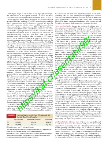
The elegant design of the PIOPED II trial highlights two impor- MRA was under 80% even when technically adequate. When tandem
tant considerations in PE diagnosis, however. The first is the critical pulmonary MRA and lower extremity MR venography were combined,
importance of performing a pretest risk assessment for PE, in order to both sensitivity and specificity were >97%, but over half of studies were
interpret diagnostic results. When compared to the composite reference technically inadequate. Thus the use of pulmonary MRA in diagnosing
51
standard, CTPA (and combined CTPA-CTV, which increases sensitivity PE should be limited to centers with pulmonary MRA expertise and only
slightly without altering specificity) performs extremely well when the for patients with a contraindication to more conventional testing.
test result is concordant with the pretest probability. For example, in a
patient deemed to have a high pretest probability for PE, the positive pre- Noninvasive Leg Studies: Because the majority of subjects with PE
dictive value of CTPA was 96%. Similarly for a patient with low pretest have detectable concomitant DVT, and up to 50% of DVT patients will
25
probability, the negative predictive value was 96%. However, for subjects have PE, the diagnostic strategy for VTE must include both entities.
with discordant test results relative to their pretest risk assessment, the Noninvasive leg studies have traditionally included impedance pleth-
https://kat.cr/user/tahir99/
predictive values drop to only 60% (Table 39-5). Thus for patients in ysmography, phleborheography, venous Dopplers, and B-mode ultra-
42
whom the clinical risk assessment is low probability for PE, a positive sound scanning of leg veins, and now extend to CT or MR venography
D-dimer test on hospital admission or an unexpected finding of right done in conjunction with pulmonary angiography. The technical details
ventricular strain on echocardiography would be useful to support the of these procedures and differences between them are beyond the scope
utility of CTPA. Conversely, if the clinical assessment for PE is high, a of this chapter, and in practice, most centers now rely upon B-mode
negative CTPA should not necessarily terminate consideration of PE. In venous ultrasonography. Venous ultrasonography is extremely helpful in
this setting, additional venous ultrasonography (if CT venography was assessing a patient with symptomatic proximal deep venous thrombosis;
not performed) or digital subtraction angiography may be warranted. in this population, the test demonstrates a sensitivity of 97%, posi-
52
The PIOPED II study also addressed the ability of CTPA to detect tive predictive value of 100%, and negative predictive value of 100%.
subsegmental PEs, as this has been a point of controversy with CTPA, Unfortunately, ultrasonography performs less well in asymptomatic
and observations were that interobserver variation became more preva- patients. Patients with symptomatic DVT are far more likely to have
53
lent with smaller clots. 45,46 It is interesting to recall that in the original a proximal than a distal, or isolated calf vein, DVT. In contrast, most
PIOPED study in 1990, subsegmental PEs accounted for 6% of the asymptomatic DVT are distal, and since fewer than half of patients
19
PEs detected, and that the interobserver agreement for pulmonary with PE have leg symptoms, it seems that PE more commonly follows
47
angiograms among these small PEs was wider than that of larger PEs asymptomatic rather than symptomatic DVT. Thus a negative venous
(66% for subsegmental PEs compared to 90% for segmental and 98% duplex should not exclude the diagnosis of PE in a patient with inter-
of lobar PEs). It would appear that subsegmental PEs are challenging mediate or high clinical risk assessment. Conversely, demonstration of
47
to diagnose even by pulmonary angiography. Furthermore, the signifi- DVT provides rationale for anticoagulation, and is accepted as evidence
42
cance of these smaller filling defects is highly uncertain. In PIOPED II, for PE if clinical suspicion is present. When anticoagulation was
the positive predictive value of a filling defect on CTPA relative to the withheld on the basis of a negative venous duplex for noncritically ill
composite PE diagnosis fell dramatically from 97% for a main or lobar patients, the rate of subsequent DVT within 6 months was low, approxi-
52
artery, to 68% for a segmental artery, to just 25% for a subsegmental mately 2%, and mortality from PE was less than 0.1%. Unfortunately,
clot. Based on retrospective review of data, some experts recommend one suspects that negative duplex results are less meaningful for criti-
42
that anticoagulation may be safely withheld for patients with exclusively cally ill patients, who retain significant risk factors for developing new
subsegmental PE when certain conditions are met. In the absence of DVT as long as they are in the ICU.
a prospective investigation, however, these recommendations seem With increasing use of peripherally inserted central catheters and
to have limited applicability to the critically ill population, given that indwelling transvenous pacemakers, deep vein thromboses in the upper
they require the patient to have adequate cardiopulmonary reserve extremities are becoming increasingly common. Prospective registries
and a transient, resolved risk factor for PE. The optimal treatment for for VTE have reported that between 5% and 10% of observed DVTs
48
12,54
subsegmental or smaller clots in the ICU remains uncertain. occur in the upper extremities, and this proportion is likely to be
Magnetic resonance angiography (MRA) technology has also pro- higher among critically ill patients, since this population is enriched for
gressed greatly in the past 2 decades, with rapidly improving resolu- malignancy, central vein catheters, and cardiac devices. Venous ultra-
tion and speed of image acquisition. MRA does not require iodinated sound appears to remain both sensitive and specific (both ~90%) in the
55
contrast, and does not have the risk of radiation associated with CTPA. setting of upper extremity DVT, despite the fact that the proximal sub-
Among initial reports in 1997, one study listed a sensitivity of 100% and clavian and brachiocephalic veins cannot be directly visualized due to
specificity of 95% when MRA was performed in 30 patients undergoing bony structures. The addition of color Doppler may reveal an abnormal
pulmonary angiography for suspected PE ; another early study reported flow pattern to suggest proximal DVT even when distal veins are pat-
49
77% sensitivity but 98% specificity. The initial optimism over MRI as a ent and compressible, though Doppler has not been proven to improve
50
55
diagnostic tool for PE has been tempered by issues of obtaining sufficient sensitivity. In critically ill patients, we advocate the extension of venous
quality scans in critically ill subjects. In the recent PIOPED III study, ultrasound to all four extremities when evaluating for PE in a patient with
25% of MRI studies were technically inadequate, and the sensitivity of risk factors for upper extremity clots, including malignancy, indwelling
central catheter or cardiac device, prolonged critical illness, hypercoagu-
lable state, or upper extremity trauma. Digital subtraction venography,
TABLE 39-5 Positive and Negative Predictive Value of CT Angiography With using intravenous contrast, is an option when venous ultrasound and
Respect to Clinical Pretest Probability CTPA are nondiagnostic, but carries much of the risk of conventional
angiography with respect to dye load and radiation exposure.
High Probability Intermediate Probability Low Probability
PPV 96 (78-99) 92 (84-96) 58 (40-73) Ventilation Perfusion (VQ) Lung Scan: V/Q scanning was traditionally
the initial test of choice in the evaluation of PE, but it has been largely
NPV 60 (32-83) 89 (82-93) 96 (92-98)
supplanted by CT scanning given that a definitive positive or negative
NPV, negative predictive value; PPV, positive predictive value. result is generally achievable in only ~20% of critically ill patients.
56
Values shown are the predictive value and (95% confidence interval). Note that CT angiography is less V/Q scans can be extremely helpful to the clinician when they provide
accurate when the CT findings are discordant with the pretest clinical probability. either a high probability result—with an attendant specificity of 85%,
Data from Kearon C, Kahn SR, Agnelli G, Goldhaber S, Raskob GE, Comerota AJ. Antithrombotic therapy ruling in the diagnosis—or a normal result, when the diagnosis of PE
47
for venous thromboembolic disease: American College of Chest Physicians Evidence-Based Clinical is virtually excluded. The frustration with V/Q scanning stems from
Practice Guidelines (8th ed). Chest. June 2008;133(suppl 6):454S-545S. the large number of tests which yield either intermediate probability
section03.indd 325 1/23/2015 2:07:35 PM