
Page 464 - Hall et al (2015) Principles of Critical Care-McGraw-Hill
P. 464
334 PART 3: Cardiovascular Disorders
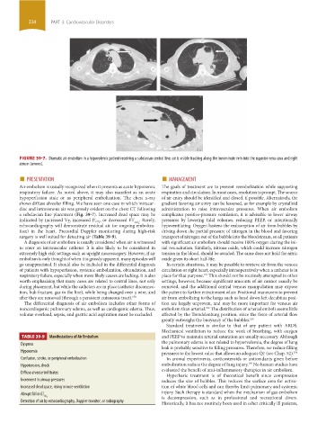
FIGURE 39-7. Dramatic air embolism in a hypovolemic patient receiving a subclavian central line; air is visible tracking along the innominate vein into the superior vena cava and right
atrium (arrows).
■ PRESENTATION ■ MANAGEMENT
Air embolism is usually recognized when it presents as acute hypoxemic The goals of treatment are to prevent reembolization while supporting
respiratory failure. As noted above, it may also manifest as an acute respiration and circulation. In most cases, resolution is prompt. The source
hypoperfusion state or as peripheral embolization. The chest x-ray of air entry should be identified and closed, if possible. Alternatively, the
shows diffuse alveolar filling. We have seen one case in which intracar- gradient favoring air entry can be lessened, as for example by crystalloid
diac and intravenous air was grossly evident on the chest CT following administration to raise intravascular pressures. When air embolism
a subclavian line placement (Fig. 39-7). Increased dead space may be complicates positive-pressure ventilation, it is advisable to lower airway
, or decreased ET . Rarely, pressures by lowering tidal volumes, reducing PEEP, or intentionally
indicated by increased Ve, increased P CO 2 CO 2
echocardiography will demonstrate residual air (or ongoing emboliza- hypoventilating. Oxygen hastens the reabsorption of air from bubbles by
tion) in the heart. Precordial Doppler monitoring during high-risk driving down the partial pressure of nitrogen in the blood and favoring
surgery is well suited for detecting air (Table 39-9). transport of nitrogen out of the bubble into the bloodstream, so all patients
A diagnosis of air embolism is usually considered when air is witnessed with significant air embolism should receive 100% oxygen during the ini-
to enter an intravascular catheter. It is also likely to be considered in tial resuscitation. Similarly, nitrous oxide, which could increase nitrogen
extremely high-risk settings such as upright neurosurgery. However, if air tension in the blood, should be avoided. The same does not hold for nitric
embolism is only thought of when it is grossly apparent, many episodes will oxide given its short half-life.
go unappreciated. It should also be included in the differential diagnosis In certain situations, it may be possible to retrieve air from the venous
of patients with hypoperfusion, systemic embolization, obtundation, and circulation or right heart, especially intraoperatively when a catheter is in
respiratory failure, especially when more likely causes are lacking. It is also place for that purpose. This should not be routinely attempted in other
191
worth emphasizing that many cases are related to central lines, not only settings, however, because significant amounts of air cannot usually be
during placement, but while the catheters are in place (catheter disconnec- removed, and the additional central venous manipulation may expose
tion, hub fracture, gas in the line), while being changed over a wire, and the patient to further entrainment of air. Positional maneuvers to prevent
after they are removed (through a persistent cutaneous tract). 190 air from embolizing to the lungs such as head down left decubitus posi-
The differential diagnosis of air embolism includes other forms of tion are largely unproven, and may be more important for venous air
noncardiogenic pulmonary edema, as well as cardiogenic edema. Thus, embolism than arterial. The distribution of arterial emboli seems little
192
volume overload, sepsis, and gastric acid aspiration must be excluded. affected by the Trendelenburg position, since the force of arterial flow
greatly outweighs the buoyancy of the bubbles. 193
Standard treatment is similar to that of any patient with ARDS.
Mechanical ventilation to reduce the work of breathing, with oxygen
TABLE 39-9 Manifestations of Air Embolism and PEEP to maintain arterial saturation are usually necessary. Although
the pulmonary edema is not related to hypervolemia, the degree of lung
Dyspnea
leak is probably sensitive to filling pressures. Therefore, we reduce filling
Hypoxemia pressures to the lowest value that allows an adequate Q ˙ t (see Chap. 52). 194
Confusion, stroke, or peripheral embolization In animal experiments, corticosteroids or antioxidants given before
195
Hypotension, shock embolization reduce the degree of lung injury. No human studies have
evaluated the benefit of anti-inflammatory therapies in air embolism.
Diffuse alveolar infiltrates
Hyperbaric treatment is of theoretical benefit since compression
Increment in airway pressures reduces the size of bubbles. This reduces the surface area for activa-
Increased dead space, rising minute ventilation tion of white blood cells and can thereby limit pulmonary and systemic
Abrupt fall in ET injury. Such therapy is standard when the mechanism of gas embolism
CO 2 is decompression, such as in professional and recreational divers.
Detection of air by echocardiography, Doppler monitor, or radiography
Historically, it has not routinely been used in other critically ill patients,
section03.indd 334 1/23/2015 2:07:39 PM