
Page 468 - Hall et al (2015) Principles of Critical Care-McGraw-Hill
P. 468
338 PART 3: Cardiovascular Disorders
specific causes identified in less than 20% of cases. Important etiologies to of the cardiac chambers. This impairment precipitates a reduction in
consider for further evaluation are those that require specific therapy, such cardiac output, followed by increases in pulmonary venous and jugular
neoplastic disorders, autoimmune disease, trauma (eg, postsurgical), and venous pressures. With inspiration, there is a fall in the driving pressure
infection (eg, tuberculosis). The vast majority of cases of acute pericarditis to fill the left ventricle, subsequently leading to a reduction in ventricu-
are viral or idiopathic, and thus are usually benign and responsive to anti- lar filling and stroke volume. The fall in left ventricular stroke volume
inflammatory drugs. For patients with a concomitant pericardial effusion, during inspiration manifests as a relative decrease in pulse pressure or
pericardiocentesis may be considered. Pericardiocentesis can assist in peak systolic pressure, which is the hallmark finding of pulsus paradoxus
diagnosis when other testing is inconclusive, and is indicated for treat- in patients with cardiac tamponade.
ment of cardiac tamponade or drug-refractory, symptomatic pericardial There are uncommon clinical presentations of cardiac tamponade.
effusions. Cardiac tamponade may be localized, when a loculated pericardial effu-
Targeted therapy of the underlying etiology of acute pericarditis sion is tactically located to impair ventricular filling. This manifestation
is indicated when a cause is identified and treatment is appropriate may occur after cardiac surgery or other postoperative settings. The
(eg, tuberculosis, uremia, thyroid disease). For analgesia and treatment loculated effusion may be present in the posterior pericardial space adja-
of the pericardial inflammation, NSAIDs are the mainstay of medical cent to the atria, which poses challenges for detection by echocardiogra-
therapy. The properties of these drugs provide effective relief in 70% phy. Posterior loculated effusions should be suspected in a postoperative
to 80% of patients. The efficacy of medical therapy varies according to patient with hemodynamic instability.
the underlying etiology, with response rates greatest among those with Low-pressure cardiac tamponade occurs without elevated jugular
idiopathic or presumed viral causes. It is important to note that relatively venous pressure because the intracardiac filling pressures are low.
6
high doses of these medications are required for them to exhibit their Examples of this manifestation are patients with tuberculosis or malig-
anti-inflammatory effect (eg, aspirin, 650-1000 mg every 6-8 hours; nancy complicated by severe dehydration. Finally, pneumopericardium
ibuprofen, 400-800 mg every 8 hours; indomethacin, 50 mg every with cardiac tamponade may result from gas-forming bacterial pericar-
8 hours). Furthermore, although these drugs can result in acute relief of ditis after penetrating chest trauma.
to reduce the risk of recurrence of inflammation. Of note, for pericarditis ■ DIAGNOSIS
symptoms, slow tapering over a period of 2 to 4 weeks is recommended
associated with myocardial infarction, NSAIDs other than aspirin should Cardiac tamponade should be suspected when there is a compatible
be avoided due to their effect of impairment of myocardial healing and history, hypotension, and an elevated jugular venous pressure or pulsus
potential for increasing the risk of mechanical complications. paradoxus. The chest x-ray (eg, “water-bottle heart”) and electrocar-
Colchicine (0.5-1.2 mg/d for 3 months), in addition to NSAIDs, is diography (eg, sinus tachycardia, electrical alternans) may be helpful.
an effective adjunctive therapy for acute pericarditis. The efficacy of Echocardiography is the primary modality for diagnosing cardiac
colchicine has been demonstrated in several randomized and retrospec- tamponade. However, knowledge of the invasive signs of tamponade
tive studies, which have shown lower rates of treatment failure (11.7% also expedites the recognition of its presence, as hemodynamic monitor-
vs 36.7%) and recurrent pericarditis (10.7% vs 32.3%) when used in ing with cardiac catheterization (ie, Swan-Ganz) is commonly available
conjunction with standard NSAID therapy. Colchicine is generally in critical care patients.
1-3
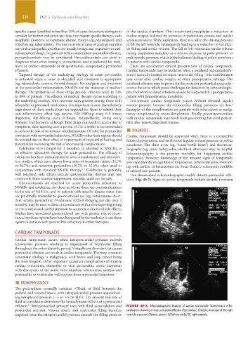
well tolerated; side effects include gastrointestinal distress and, less Two-dimensional echocardiography readily detects pericardial effu-
commonly, bone marrow suppression, myositis, and liver toxicity. sions (Fig. 40-2). Signs of cardiac tamponade include diastolic inversion
Glucocorticoids are reserved for acute pericarditis refractory to
NSAIDs and colchicine, for those in whom there are contraindications
to the use of NSAIDs, and in patients with specific disease states that
are potentially amenable to glucocorticoid use (eg, autoimmune disor-
ders, uremic pericarditis). Prednisone (0.25-0.50 mg/kg per day over 3
months) may be used in these circumstances with a slow taper beginning
at 2 to 4 weeks and careful attention to occurrence of steroid side effects.
Studies have associated glucocorticoid use with greater risk of recur-
rence, but these reports have been hampered by the tendency to use these
agents in patients with pericarditis refractory to other therapies.
CARDIAC TAMPONADE
Cardiac tamponade occurs when intrapericardial pressure exceeds
intracardiac pressure, resulting in impairment of ventricular filling
throughout the entire diastolic period. Virtually any disorder that causes
pericardial effusion can result in cardiac tamponade. The most common
atraumatic etiology is malignancy, with breast and lung cancer being
the most frequent. Other important causes are complications of invasive
cardiac procedures, idiopathic or viral pericarditis, aortic dissection
with disruption of the aortic valve annulus, tuberculosis, uremia, and
pericarditis or ventricular wall rupture from myocardial infarction.
■ PATHOPHYSIOLOGY
The pericardium normally contains <50 mL of fluid between the
parietal and visceral layers, with intrapericardial pressure approximat-
ing intrapleural pressure (−5 to +5 cm H O). The amount and rate of
2
fluid accumulation determine the hemodynamic effects of a pericardial
effusion. Intrapericardial pressure rises with fluid accumulation and FIGURE 40-2. Echocardiographic features of cardiac tamponade. Transthoracic echo-
4,5
pericardial restraint. Venous return and ventricular filling becomes cardiogram showing a large pericardial effusion (Top, arrows). Diastolic inversion of the right
impaired once the intrapericardial pressure exceeds the filling pressure ventricle is present (Bottom, arrow). LV, left ventricle; RV, right ventricle.
section03.indd 338 1/23/2015 2:07:41 PM