
Page 465 - Hall et al (2015) Principles of Critical Care-McGraw-Hill
P. 465
CHAPTER 39: Pulmonary Embolic Disorders: Thrombus, Air, and Fat 335
in large part due to the availability of decompression chambers and endothelium, their concentration in the systemic circulation during
the risks attendant with transferring critically ill patients. In France, critical illness does not rise sufficiently to account for lung injury.
the sole decompression unit serving Paris published their experience
of decompressing 119 patients with iatrogenic air embolism over an Traumatic Embolism: Fracture of bone releases neutral fat which
11-year period, and reported a 1-year mortality rate of 21%. The only embolizes into the pulmonary vasculature. The derivation of this fat
196
reported adverse outcome was one seizure during hyperbaric treat- from bone is supported by the finding of coincident particles of bone
ment, which resolved upon decreasing the fraction of inspired oxygen. marrow at autopsy in patients with long bone fractures and by echo-
The hyperbaric protocol used consisted of one dive, with 15 minutes at cardiographic studies showing frequent, often dramatic embolism at
201
4 atmospheres (ATA), then a 45-minute plateau at 3 ATA, followed by the time of medullary reaming. Even in traumatic embolism, the
45 minutes at 2 ATA. Since patients usually respond readily to stan- syndrome appears to be more than a localized, mechanical obstruc-
196
dard supportive measures, and since the syndrome typically resolves in tion, however. Intravascular hydrolysis of fat by lung lipase releases
only 24 to 48 hours, we have not typically utilized hyperbaric therapy toxic FFAs, which generate endothelial injury. Systemic findings
except in the most extreme and persistent cases. While some advocate in FES probably relate to passage of venous fat emboli across the
that hyperbaric therapy must be initiated early to have an effect, we pulmonary circulation, although serum-derived fat may play a role.
would stress the importance of resuscitation prior to leaving the ICU for Elevated right heart pressures following embolism may open a probe-
202
an unproven therapy. When such patients are transported by air, a pres- patent foramen ovale, causing severe, even fatal systemic embolism.
surized craft flying at low altitude should be requested. Further, fat can cross the pulmonary circuit even in the absence of a
right-to-left shunt, as has been shown in experimental animals. Fat
was able to traverse the pulmonary microcirculation even though
FAT EMBOLISM 15-micron radiolabeled microspheres could not, perhaps due to
enhanced deformability of fat emboli. 203
The fat embolism syndrome (FES) is associated with fat particles in the
microcirculation of the lung. It consists typically of lung dysfunction, ■ CLINICAL MANIFESTATIONS
neurologic manifestations, and petechiae, usually following a latent
interval. It is most common following long bone fractures, typically Following injury, there is usually a latent interval of 12 to 72 hours
presenting as dyspnea and confusion. However, FES is seen after other before the syndrome becomes evident. The dominant findings are
forms of trauma and in several nontraumatic conditions as well. For related to lung injury and neurologic dysfunction. Patients with FES
example, FES has been proposed as a major cause of the acute chest present as ARDS, with dyspnea, hypoxemia, and a diffuse lung lesion.
syndrome in patients with sickle cell disease (see Chap. 96). More In addition, there is often confusion, obtundation, or coma, signs due
197
recently, patients have been presenting with FES following often unregu- to cerebral fat embolism rather than coincident hypoxemia. The typical
lated cosmetic procedures involving silicone or mineral oil injection. neuropathologic findings include fat microemboli and diffuse petechial
198
After long bone or pelvic fracture, the incidence of the syndrome is at hemorrhagic infarcts. Petechiae are also seen on the skin, particularly
least 10% when patients are prospectively screened, although serious over the upper chest, neck, and face, though they appear only in 50% of
199
clinical manifestations are seen in only 1% to 3%. Since the clinical patients. On fundoscopic examination, embolized fat may be detected
presentation is usually mild, FES is often unrecognized. Even when lung in retinal vessels (Purtscher retinopathy). Often thrombocytopenia and
injury is obvious, its cause may be attributed to infection, aspiration, or anemia are present. Rare patients will develop a full blown acute right
traumatic ARDS, rather than to fat embolization. Some of the causes of heart syndrome.
The diagnosis is usually based on the clinical findings in a patient at risk
FES are presented in Table 39-10. for FES. Fat globules in the urine are neither sensitive nor specific for the
■ PATHOPHYSIOLOGY diagnosis of FES. Attempts have been made to find alternative, objective
Nontraumatic Embolism: Fat globules are seen in pulmonary (and other) means of diagnosis, since this might be useful in devising prophylactic
or therapeutic strategies. Fat can be detected in bronchoalveolar lavage
vessels at autopsy and can be found in venous blood. In contrast to specimens in many patients following trauma, but this finding appears not
traumatic embolism, the fat is probably not derived from bone marrow, to be a reliable means of diagnosis. Fat can also be seen in spun samples
204
but rather arises from lipids in the blood. Serum from acutely ill patients of blood withdrawn from a wedged pulmonary artery catheter, but this
has the capacity to agglutinate chylomicrons and very low-density lipo- finding too, is neither sensitive nor specific. 205
proteins (VLDL), as well as liposomes of nutritional fat emulsions.
200
It has been proposed that C-reactive protein (CRP), which provokes ■
the calcium-dependent agglutination of each of these lipid-containing PROPHYLAXIS AND TREATMENT
substances, may underlie nontraumatic FES. Since CRP is dramatically Because patients with long bone fracture have such a well-defined risk
elevated in trauma, sepsis, and inflammatory disorders, this provides factor for FES, prophylactic strategies have been evaluated. The least
a mechanism for fat embolization. An alternative, but less attractive, controversial strategy has involved a shift toward early fixation of long
hypothesis implicates the liberation of free fatty acids (FFAs) from fat bone fractures, even in patients with multiple trauma. Early fixation
stores. Although FFAs are known to injure the pulmonary vascular decreases the incidence of FES, as well as of ARDS and pneumonia, and
reduces length of stay. 206-208 Orthopedic surgery, and particularly total
hip arthroplasty, carries a high risk of fat embolism, which has been
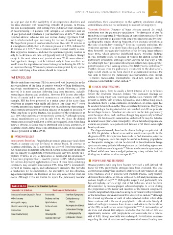
TABLE 39-10 Causes of Fat Embolism Syndrome demonstrated by transesophageal echocardiography to occur during
the preparation of the femur and insertion of the femoral component.
Traumatic Fat Embolism Nontraumatic Fat Embolism
Specific surgical techniques such as using bone vacuum with preparation
Long bone fracture (especially femur) Pancreatitis Fatty liver of pregnancy of the femur have shown decreased TEE-detected fat emboli, although
209
Other fractures Diabetes mellitus Cardiopulmonary the clinical significance of such of echo-detected emboli is less clear.
Orthopedic surgery Lipid infusions bypass More controversial is the use of prophylactic corticosteroids. Nearly all
trials of methylprednisolone have shown a reduction in the incidence
Blunt trauma to fatty organs (liver) Sickle cell crisis Decompression sickness of the FES, as well as less severe hypoxemia. 210-212 A meta- analysis of
Liposuction Burns Corticosteroid therapy sis trials and almost 400 subjects concluded that the risk of FES was
Bone marrow biopsy Osteomyelitis Lymphangiography significantly reduced with prophylactic corticosteroids, for a relative
risk of 0.22, though mortality was unchanged. Nevertheless, concerns
Alcoholic fatty liver Cyclosporine infusion
regarding the risk of infection and impairment of wound healing have
section03.indd 335 1/23/2015 2:07:39 PM