
Page 477 - Hall et al (2015) Principles of Critical Care-McGraw-Hill
P. 477
CHAPTER 41: Valvular Heart Disease 347
(increased afterload due to increased wall stress and systolic pressure).
• Acute presentation is usually with dyspnea, less common with The left ventricle dilates progressively, with both eccentric and concentric
syncope or angina. hypertrophy. The increased diastolic volume allows for augmentation of
• Harsh, loud, midsystolic murmur and decreased S2 are typical of stroke volume (Frank-Starling mechanism), and maintains cardiac out-
severe AS. This may be reduced or absent in patients with reduced put in the normal range for many years despite presence of severe AR.
ejection fraction. Once compensatory mechanisms are overwhelmed, the disease tends to
• Echocardiography is the cornerstone of diagnosis. Valve area progress rapidly. Left ventricular volumes (left ventricular end-systolic
diameter >50 mm, left ventricular end-diastolic diameter >70 mm) can
<1.0 cm and gradient ≥40 mm Hg are diagnostic of severe AS. be used to predict which patients are more likely to have progressive
2
• Low output/low gradient AS can be diagnosed with low-dose dobu- disease and the development of left ventricular failure. Indexed left
tamine stress. Presence of contractile reserve predicts good outcome. ventricular dimensions are better predictors in patients of small body
• Cardiac catheterization verifies severity of AS in difficult cases and size and in women. 21
provides preoperative coronary angiography. In acute onset AR, the left ventricle has to accommodate suddenly a
• Medical management is only temporizing. large regurgitant volume. Left ventricular dilation is limited by the com-
pliance of the ventricle and by the constraining pericardium. As such,
• Temporizing aortic balloon valvuloplasty may be used. small increase in regurgitant volume may lead to a dramatic increase
• Surgical or transcatheter aortic valve replacement are the only in left ventricular diastolic pressure. This leads to an increase in left
treatments with long-term success. Decision should be made by atrial pressure, causing pulmonary congestion/edema. The combina-
the heart team (surgeon and cardiologist). tion of decreased aortic diastolic pressure and increased left ventricular
diastolic pressure leads to a dramatic decrease in coronary perfusion
pressure. Acute AR is especially difficult to tolerate by patients with
a very stiff left ventricle due to preexisting concentric hypertrophy;
AORTIC REGURGITATION this entity is now more commonly present with a resurgence of aortic
■ ETIOLOGY balloon valvuloplasty (postdilatation AR) and increasing use of TAVR
(periprosthetic AR).
The presentation and management of patients with severe aortic regur-
gitation (AR) depends on the nature of underlying disease. Acute severe ■ CLINICAL PRESENTATION
AR is rare, but is a true medical and surgical emergency. It can be the Patients with decompensation of chronic long-standing AR have the
result of endocarditis (leaflet or annular destruction), aortic dissec- classical features of AR in addition to the signs and symptoms of
tion (compromised leaflet coaptation), or traumatic (leaflet or annular the acute decompensated state. The heart is enlarged, with displaced
tear/rupture from blunt chest trauma or aortic balloon valvuloplasty; apical impulse. The heart sounds are usually normal (unless pulmonary
the aortic valve is the most commonly involved valve in blunt chest hypertension is present). There is a soft diastolic decrescendo murmur
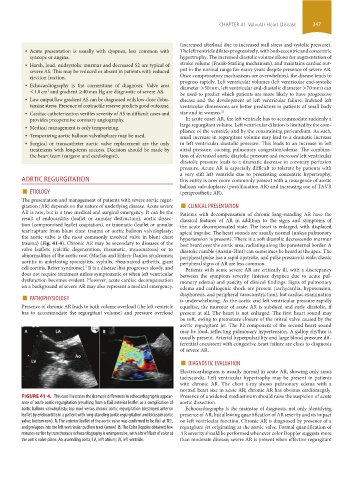
trauma) (Fig. 41-4). Chronic AR may be secondary to diseases of the best heard over the aortic area, radiating along the parasternal border. A
valve leaflets (calcific degeneration, rheumatic, myxomatous) or to diastolic rumble (Austin-Flint) can sometimes be heard at the apex. The
abnormalities of the aortic root (Marfan and Ehlers-Danlos syndromes; peripheral pulse has a rapid upstroke, and pulse pressure is wide; classic
aortitis in ankylosing spondylitis, syphilis, rheumatoid arthritis, giant peripheral signs of AR are less common.
cell aortitis, Reiter syndrome). It is a disease that progresses slowly, and Patients with acute severe AR are critically ill, with a discrepancy
5
does not require treatment unless symptomatic or when left ventricular between the symptom severity (intense dyspnea due to acute pul-
dysfunction becomes evident. However, acute cardiac decompensation monary edema) and paucity of clinical findings. Signs of pulmonary
on a background of severe AR may also represent a medical emergency. edema and cardiogenic shock are present (tachycardia, hypotension,
■ PATHOPHYSIOLOGY diaphoresis, and peripheral vasoconstriction), but cardiac examination
is underwhelming. As the aortic and left ventricular pressure rapidly
Presence of chronic AR leads to both volume overload (the left ventricle equalize, the murmur of acute AR is subdued and early diastolic, if
has to accommodate the regurgitant volume) and pressure overload present at all. The heart is not enlarged. The first heart sound may
be soft, owing to premature closure of the mitral valve caused by the
aortic regurgitant jet. The P2 component of the second heart sound
may be loud, reflecting pulmonary hypertension. A gallop rhythm is
usually present. Arterial hyperpulsatility and large blood pressure dif-
ferential coexistent with congestive heart failure are clues to diagnosis
of severe AR.
■ DIAGNOSTIC EVALUATION
Electrocardiogram is usually normal in acute AR, showing only sinus
tachycardia. Left ventricular hypertrophy may be present in patients
with chronic AR. The chest x-ray shows pulmonary edema with a
normal heart size in acute AR; chronic AR has obvious cardiomegaly.
FIGURE 41-4. This case illustrates the dramatic differences in echocardiographic appear- Presence of a widened mediastinum should raise the suspicion of acute
ance of acute aortic regurgitation (resulting from a flail anterior leaflet as a complication of aortic dissection.
aortic balloon valvuloplasty; top row) versus chronic aortic regurgitation (destroyed anterior Echocardiography is the mainstay of diagnosis, not only identifying
leaflet by endocarditis in a patient with long-standing aortic regurgitation and bicuspid aortic presence of AR, but allowing quantification of AR severity and its impact
valve; bottom row). A. The anterior leaflet of the aortic valve was confirmed to be flail at TEE, on left ventricular function. Chronic AR is diagnosed by presence of a
and prolapses into the left ventricular outflow tract (arrow). B. The Color Doppler obtained few regurgitant jet originating at the aortic valve. Formal quantification of
minutes earlier by transthoracic echocardiography is unimpressive, with a brief flash of color at AR severity should be performed whenever color Doppler suggests more
the aortic valve plane. Ao, ascending aorta; LA, left atrium; LV, left ventricle. than moderate disease; severe AR is present when effective regurgitant
section03.indd 347 1/23/2015 2:07:50 PM