
Page 489 - Hall et al (2015) Principles of Critical Care-McGraw-Hill
P. 489
CHAPTER 42: Aortic Dissection 359
2 3
1
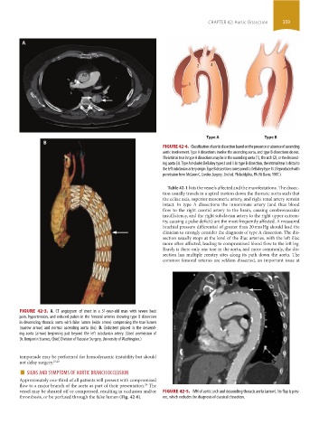
Type A Type B
FIGURE 42-4. Classification of aortic dissection based on the presence or absence of ascending
aortic involvement. Type A dissections involve the ascending aorta, and type B dissections do not.
The intimal tear in type A dissections may be in the ascending aorta (1), the arch (2), or the descend-
ing aorta (3). Type A includes DeBakey types I and II. In type B dissection, the intimal tear is distal to
the left subclavian artery origin. Type B dissections correspond to DeBakey type III. (Reproduced with
permission from McGoon C. Cardiac Surgery. 2nd ed. Philadelphia, PA: FA Davis; 1987.)
Table 42-1 lists the vessels affected and the manifestations. The dissec-
tion usually travels in a spiral motion down the thoracic aorta such that
the celiac axis, superior mesenteric artery, and right renal artery remain
intact. In type A dissections the innominate artery (and thus blood
flow to the right carotid artery to the brain, causing cerebrovascular
insufficiency, and the right subclavian artery to the right upper extrem-
ity, causing a pulse deficit) are the most frequently affected. A measured
brachial pressure differential of greater than 20 mm Hg should lead the
clinician to strongly consider the diagnosis of type A dissection. The dis-
section usually stops at the level of the iliac arteries, with the left iliac
more often affected, leading to compromised blood flow to the left leg.
Rarely is there only one tear in the aorta, and more commonly, the dis-
section has multiple reentry sites along its path down the aorta. The
common femoral arteries are seldom dissected, an important issue at
FIGURE 42-3. A. CT angiogram of chest in a 51-year-old man with severe back
pain, hypertension, and reduced pulses in the femoral arteries showing type B dissection
in descending thoracic aorta with false lumen (wide arrow) compressing the true lumen
(narrow arrow) and normal ascending aorta (Ao). B. Endostent placed in the descend-
ing aorta (arrow) beginning just beyond the left subclavian artery. (Used permission of
Dr. Benjamin Starnes, Chief, Division of Vascular Surgery, University of Washington.)
tamponade may be performed for hemodynamic instability but should
not delay surgery. 27,28
■ SIGNS AND SYMPTOMS OF AORTIC BRANCH OCCLUSION
Approximately one-third of all patients will present with compromised
29
flow to a major branch of the aorta as part of their presentation. The
vessel may be sheared off or compressed, resulting in occlusion and/or FIGURE 42-5. IMH of aortic arch and descending thoracic aorta (arrow). No flap is pres-
thrombosis, or be perfused through the false lumen (Fig. 42-6). ent, which excludes the diagnosis of classical dissection.
section03.indd 359 1/23/2015 2:08:20 PM