
Page 592 - Hall et al (2015) Principles of Critical Care-McGraw-Hill
P. 592
412 PART 4: Pulmonary Disorders
It is easiest to derive clinically useful information about the patient’s Ppeak
respiratory system when volume-preset modes such as ACV or SIMV b x Pplat
are used. At least when the patient is passive, the pressure at the air-
way opening (Pao) and the pressure versus time waveform reflect the Pao
mechanical properties of the respiratory system, yielding valuable a a
clinical information. During pressure-preset modes, such as pressure- PEEP
support ventilation (PSV) and pressure-control ventilation (PCV), some
information can be derived from the flow versus time waveform, but
this information is generally less readily interpreted than that obtained
during volume-preset ventilation. Below we review the determinants Pelast
of the pressure and flow versus time waveforms during volume-preset,
then pressure-preset, ventilation, including how to recognize and quan-
titate autoPEEP as well as a method for using this information to adjust
the ventilator. Volume-pressure loops are reviewed in terms of how Presist
they may aid management of the patient with acute lung injury (ALI)
or acute respiratory distress syndrome (ARDS) but we also review the PEEP
simpler use of the stress index for this same purpose. The potentially
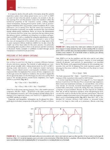
confounding effect of patient effort on the pressure and flow waveforms FIGURE 48-1. During constant flow, volume-preset ventilation of a passive patient,
is discussed. Finally, examples of problems revealed through careful Pao is composed of resistive and elastic elements, the latter consisting of the end-expiratory
interpretation of waveforms are presented. pressure (PEEP or autoPEEP) and a component proportional to the change in volume and the
respiratory system compliance. The second breath includes an inspiratory pause allowing
determination of the components of Pao.
PRESSURE AT THE AIRWAY OPENING
■ VOLUME-PRESET MODES First, PEEP is set on the ventilator and this value can be used when
autoPEEP is absent. AutoPEEP is present, however, in most ventilated
Gas is driven to and from the lung by a pressure difference between critically ill patients, and methods for quantitating it are described
2
alveolus and airway opening. The majority of adult patients are venti- below. The Ppeak can be apportioned between its two remaining com-
lated, at least initially, with a volume-preset mode (ie, ACV or IMV), ponents, Pres and Pel, by stopping flow (end-inspiratory pause ) and
1
3
allowing ready determination of the respiratory system mechanics. allowing the Pres term to fall to 0. When flow is 0, Pao drops to a lower
When a muscle-relaxed patient is mechanically ventilated at constant Pplat. Then:
inspiratory flow, the inspiratory Pao consists of three components: one
to drive gas across the inspiratory resistance, the second to expand the Pres = Ppeak – Pplat
alveoli against the elastic recoil of the lungs and chest wall, and the third The final component (Pel = Pplat − Total PEEP) is proportional to the
equal to the alveolar pressure present before inspiratory flow begins elastance of the respiratory system and the tidal volume.
(PEEP or autoPEEP) (Fig. 48-1). At normal inspiratory flow rates in the range of 1 L/s, Pres is typically
Pao = Pres + Pel + Total PEEP or between 4 and 10 cm H O. Elevated Pres is found with high inspiratory
2
flow or increased inspiratory resistance. At constant flow, a rise in Pres
may indicate, for example, increased bronchospasm or partial endo-
Pao = Flow × Rrs + DV × Ers + Total PEEP
I tracheal tube obstruction. Conversely, falling Pres may correspond to
where Pao is the airway opening pressure, Pres is the resistive pressure a response to bronchodilators. Because the Pres depends on ventilator
component, Pel (Pel = Pplat − Total PEEP) is the elastic pressure term, flow rate, as well as inspiratory resistance, when interpreting its value,
Rrs is inspiratory resistance, ΔV is the increment in lung volume, Ers is one must be careful to take the set flow rate into consideration. The most
elastance of the respiratory system, and Total PEEP is applied PEEP or dramatic example of potential error in this regard is when the inspiratory
autoPEEP, whichever is higher. flow is set to a decelerating profile (Fig. 48-2). Since Pel = ΔV × Ers,
Diagnostic and therapeutic information can be gleaned by distin- elevated Pel indicates excessive tidal volume or increased elastic
guishing the individual components of the peak Pao (Ppeak), as follows. recoil of the lungs or chest wall, as in pulmonary fibrosis, acute lung
60 A B
Flow
(lpm)
−60
40
Pao
(cm H O)
2
5
FIGURE 48-2. This is a passive patient with modest airflow obstruction ventilated with a volume-preset mode and square wave flow (panel A) at 60 lpm or decelerating flow (panel B)
beginning at 60 lpm. A 0.4-second end-inspiratory pause is set in order to allow determination of Pplat. Notice that there is a significant difference between Ppeak and Pplat (40-22) during
square wave ventilation but not during decelerating flow (27-22) because flow is so low during the later parts of the breath.
section04.indd 412 1/23/2015 2:19:04 PM