
Page 595 - Hall et al (2015) Principles of Critical Care-McGraw-Hill
P. 595
CHAPTER 48: Ventilator Waveforms: Clinical Interpretation 415
alveolar pressure equals the ventilator P . Flow waveform displays facili- impede venous return, heighten the risk of barotrauma, and improve
I
tate adjustments of T and respiratory rate as discussed below. oxygenation. In addition, autoPEEP increases the work of breathing
I
When patients are breathing spontaneously on PSV, thereby deter- and impairs the patient’s ability to trigger the ventilator. For these rea-
mining their own T and rate, waveform analysis aids the identification sons, it is imperative to monitor routinely the presence and amount of
I
of patient-ventilator asynchrony. This may be especially important autoPEEP in mechanically ventilated patients.
during noninvasive ventilation (NIV). Common problems during NIV AutoPEEP is present when the expiratory flow tracing reveals persis-
include failure of the ventilator to recognize the onset of patient inspira- tent end-expiratory flow (see Fig. 48-10). Additionally, when the Pres
tion (generally due to autoPEEP, as described below) and excessive T I is lower than the height of the early step change in the Pao waveform,
(related to respiratory mechanics and the threshold at which inspiratory autoPEEP is likely to be present. Several methods for quantifying
pressure switches off). 5 autoPEEP are available, but the one typically used clinically is the end-
expiratory port occlusion method. Modern ventilators facilitate this
6
Volume-Preset Modes: During ACV and SIMV, flow is set by the physician determination by providing an expiratory pause function. This method
either directly or indirectly through the choice of minute ventilation and will not provide accurate estimation of autoPEEP if there is a leak in the
rate. Flow may also be altered by changes in rise time, inspiratory plateau, tubing or around the endotracheal tube cuff, there is gas flow into the
inspiratory to expiratory ratios, and other settings, depending on the circuit during expiration (as during continuous nebulization of broncho-
particular ventilator in use. Flow waveforms can reveal the effects on flow dilators), or the patient is not fully passive during the maneuver. In one
of other setting changes, as discussed below, and also whether the survey of ventilated patients, quantitation of autoPEEP was possible by
flow profile (square, decelerating, or sine) has been inadvertently changed.
the end-expiratory port occlusion technique in only one-third, because
patient effort confounded the airway pressure. Serial measurement
2
THE OBSTRUCTED PATIENT of autoPEEP may give information regarding the obstructed patient’s
■ CLUES IN THE WAVEFORMS response to bronchodilator therapy (if minute ventilation is constant). 9
The waveform indications of increased respiratory resistance are Using Peep to Ease Triggering: AutoPEEP presents an inspiratory thresh-
old load to the spontaneously breathing patient, as discussed in Chap. 54.
10
(1) increased Pres when an end-inspiratory pause has been set; (2) a high
shoulder on the early portion of the Pao versus time tracing (Fig. 48-9); The work of breathing due to this inspiratory threshold load is roughly
equal in magnitude to the excessive resistive work of breathing in
(3) low and prolonged expiratory flow, often with persistent flow at end- 11
expiration (Fig. 48-10); (4) the presence of two components to the expi- patients with COPD exacerbations, contributing to distress even when
on the ventilator. Thus therapy should be directed at reducing autoPEEP
ratory waveform (indicating early airway collapse as in Fig. 48-6); and
(5) scooping of the expiratory flow-volume curve. Significant increases when it is present. Meanwhile, PEEP can be applied externally, greatly
easing the effort required to trigger the breath. In intubated patients
in airway resistance are often associated with the presence of autoPEEP,
especially when large minute ventilations are given, as described below. with acute-on-chronic respiratory failure, continuous positive airway
pressure (CPAP) has been demonstrated to reduce the work of breathing
Determining AutoPEEP: The autoPEEP effect occurs when there is insuf- by nearly 50%. In patients with COPD and acute respiratory failure,
12
ficient time for the respiratory system to return to functional residual nasal CPAP immediately improves respiratory rate, sensation of dys-
capacity by end-expiration. Short expiratory times, high minute pnea, and the P CO 2 . The amount of autoPEEP is largely independent
13
6,7
volumes, and increased expiratory resistance contribute to autoPEEP, of the set PEEP, since the airways of obstructed patients behave more
but all of these need not be present. AutoPEEP is present in the majority like Starling resistors than like Ohmic resistors, much as the rate of flow
of ventilated patients with asthma and COPD (and in many during spon- of water over a waterfall is unrelated to how far the water will fall into
taneous breathing), but is also seen in ARDS and other settings with the pool below (Fig. 48-11). As long as the set PEEP is not higher than
8
high minute ventilations. In many regards autoPEEP acts like PEEP to
2
l/s Flow-time s
1.1
Flow-time Flow-time
0 6
−1.1
cm H O Pressure-time s
2
50
Pressure-time Pressure-time
0 6
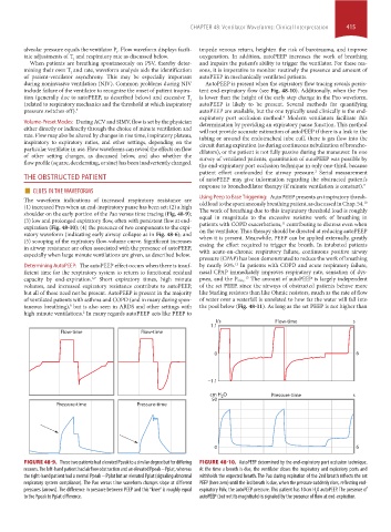
FIGURE 48-9. These two patients had elevated Ppeak to a similar degree but for differing FIGURE 48-10. AutoPEEP determined by the end-expiratory port occlusion technique.
reasons. The left-hand patient had airflow obstruction and an elevated Ppeak – Pplat, whereas At the time a breath is due, the ventilator closes the inspiratory and expiratory ports and
the right-hand patient had a normal Ppeak – Pplat but an elevated Pplat (signaling abnormal withholds the expected breath. The Pao during expiration of the 2nd breath reflects the set
respiratory system compliance). The Pao versus time waveform changes slope at different PEEP (here zero) until the 3rd breath is due, when the pressure suddenly rises, reflecting end-
pressures (arrows). The difference in pressure between PEEP and this “knee” is roughly equal expiratory Palv, the autoPEEP pressure. This patient has 10 cm H O autoPEEP. The presence of
2
to the Ppeak to Pplat difference. autoPEEP (but not its magnitude) is signaled by the presence of flow at end-expiration.
section04.indd 415 1/23/2015 2:19:08 PM