
Page 607 - Hall et al (2015) Principles of Critical Care-McGraw-Hill
P. 607
CHAPTER 49: Management of the Ventilated Patient 427
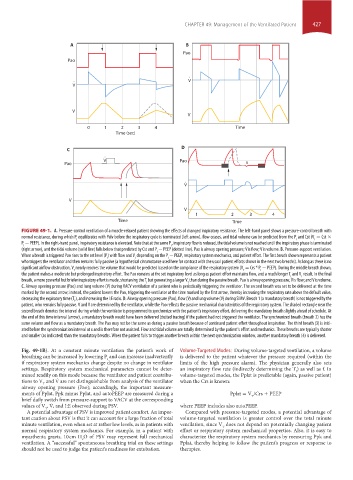
A B
Pao
Pao
.
. V
V
V
V
0 1 2 3 4 Time
Time (sec)
D
C
.
V Pao
Pao x
.
V
V
V
1 2 3 4
Time Time
FIGURE 49-1. A. Pressure-control ventilation of a muscle-relaxed patient showing the effects of changed inspiratory resistance. The left-hand panel shows a pressure-control breath with
normal resistance, during which P equilibrates with Palv before the inspiratory cycle is terminated (left arrow), flow ceases, and tidal volume can be predicted from the P and Cst (V = Cst ×
T
I
I
P − PEEP). In the right-hand panel, inspiratory resistance is elevated. Note that at the same P, inspiratory flow is reduced, the tidal volume is not reached until the inspiratory phase is terminated
I
I
(right arrow), and the tidal volume (solid line) falls below that predicted by Cst and P − PEEP (dotted line). Pao is airway opening pressure; V ˙ is flow; V is volume. B. Pressure-support ventilation.
I
When a breath is triggered Pao rises to the set level (P) with flow and V depending on the P − PEEP, respiratory system mechanics, and patient effort. The first breath shown represents a patient
T
I
I
who triggers the ventilator and then remains fully passive (a hypothetical circumstance used here for contrast with the usual patient efforts shown in the next two breaths). As long as there is no
significant airflow obstruction, V nearly reaches the volume that would be predicted based on the compliance of the respiratory system (V = Crs * P − PEEP). During the middle breath shown,
T
T
I
the patient makes a moderate but prolonged inspiratory effort. The Pao remains at the set inspiratory level as long as patient effort maintains flow, and a much longer T and V result. In the final
T
I
breath, a more powerful but briefer inspiratory effort is made, shortening the T but generating a larger V than during the passive breath. Pao is airway opening pressure; V ˙ is flow; and V is volume.
T
I
C. Airway opening pressure (Pao) and lung volume (V) during VACV ventilation of a patient who is periodically triggering the ventilator. The second breath was set to be delivered at the time
marked by the second arrow; instead, the patient lowers the Pao, triggering the ventilator at the time marked by the first arrow, thereby increasing the respiratory rate above the default value,
decreasing the expiratory time (T ), and increasing the I:E ratio. D. Airway opening pressure (Pao), flow (V ˙ ) and lung volume (V) during SIMV. Breath 1 (a mandatory breath) is not triggered by the
E
patient, who remains fully passive. V and V ˙ are determined by the ventilator, while the Pao reflects the passive mechanical characteristics of the respiratory system. The shaded rectangle near the
second breath denotes the interval during which the ventilator is programmed to synchronize with the patient’s inspiratory effort, delivering the mandatory breath slightly ahead of schedule. At
the end of this time interval (arrow), a mandatory breath would have been delivered (dotted tracing) if the patient had not triggered the ventilator. The synchronized breath (breath 2) has the
same volume and flow as a mandatory breath. The Pao may not be the same as during a passive breath because of continued patient effort throughout inspiration. The third breath (3) is initi-
ated before the synchronization interval at x and is therefore not assisted. Flow and tidal volume are totally determined by the patient’s effort and mechanics. These breaths are typically shorter
and smaller (as indicated) than the mandatory breaths. When the patient fails to trigger another breath within the next synchronization window, another mandatory breath (4) is delivered.
Fig. 49-1B). At a constant minute ventilation the patient’s work of Volume-Targeted Modes: During volume-targeted ventilation, a volume
breathing can be increased by lowering P and can increase inadvertently is delivered to the patient whatever the pressure required (within the
I
if respiratory system mechanics change despite no change in ventilator limits of the high pressure alarm). The physician generally also sets
settings. Respiratory system mechanical parameters cannot be deter- an inspiratory flow rate (indirectly determining the T ) as well as f. In
I
mined readily on this mode because the ventilator and patient contribu- volume-targeted modes, the Pplat is predictable (again, passive patient)
tions to V and V ˙ are not distinguishable from analysis of the ventilator when the Crs is known:
T
airway opening pressure (Pao); accordingly, the important measure-
ments of Pplat, Ppk minus Pplat, and autoPEEP are measured during a Pplat = V /Crs + PEEP
brief daily switch from pressure-support to VACV at the corresponding T
values of V , V ˙ , and I:E observed during PSV. where PEEP includes also autoPEEP.
T
A potential advantage of PSV is improved patient comfort. An impor- Compared with pressure-targeted modes, a potential advantage of
tant caution about PSV is that it can account for a large fraction of total volume-targeted ventilation is greater control over the total minute
minute ventilation, even when set at rather low levels, as in patients with ventilation, since V does not depend on potentially changing patient
T
normal respiratory system mechanics. For example, in a patient with effort or respiratory system mechanical properties. Also, it is easy to
myasthenia gravis, 10 cm H O of PSV may represent full mechanical characterize the respiratory system mechanics by measuring Ppk and
2
ventilation. A “successful” spontaneous breathing trial on these settings Pplat, thereby helping to follow the patient’s progress or response to
should not be used to judge the patient’s readiness for extubation. therapies.
section04.indd 427 1/23/2015 2:19:18 PM