Page 602 - Hall et al (2015) Principles of Critical Care-McGraw-Hill
P. 602

422     PART 4: Pulmonary Disorders


                                                     30

                                                     20


                                                     10

                                                     0
                                                     .5
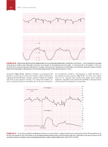
                 FIGURE 48-26.  Wedge pressure (Ppw) tracing in this ventilated patient who was making strong respiratory efforts. Since the Ppw varies from 20 to −5 cm H O, reading the tracing properly
                                                                                                          2
                 (at end-expiration) is vitally important. If this patient were passive, one would expect the end-expiratory pressure to be roughly −5 cm H O, but then the I:E ratio would be 2:1 (inverse ratio
                                                                                              2
                 ventilation), the fall in pressure during (presumed) expiration is too abrupt, and the (presumed) inspiratory pressure should continue to rise (rather than plateauing) throughout the breath.
                 Instead, this ventilated patient is quite active and pulls the Ppw down during inspiration. The end-expiratory pressure is 15 cm H O.
                                                                                        2
                 discussed in Figure 48-26. Additional confidence can be gained at the   the hemodynamic waveform. End-expiration is readily identified on
                 bedside by examining the patient and ventilator while simultaneously   the ventilator pressure waveform (Fig. 48-27). One then moves 200 ms
                 displaying the hemodynamic tracing in question. Perhaps the simplest   earlier on the timescale (since patient effort begins before machine
                 and most accurate approach, however, is to connect the ventilator cir-  inspiration, especially in the patient with autoPEEP, as discussed above)
                 cuit to a pressure transducer and display this on the same timescale as   and measures the hemodynamic pressure there.


                                                                                                     40
                                                          10 mmHg/cm
                                                                                                     35
                                            1 sec                                                    30
                                                                                                     25
                                                                                                     20
                                                                                                     CVP
                                                                                                     10
                                                                                                     5
                                                                                                     0
                                                                                                     −5
                                                                                                     −10
                                                          10 mmHg/cm                                 35
                                                                                                     30
                                                                                                     25
                                                                                                     20
                                                                                                     PRS
                                                                                                     10
                                                                                                     5
                                                                                                     0
                                                       200 msec
                                                                                                     −5

                 FIGURE 48-27.  The least equivocal method for identifying end-expiration on a vascular waveform is to display simultaneously the vascular pressure (here the CVP; top waveform) and the
                 Pao (PRS; bottom waveform). A point 200 ms before Pao rises (signaling inspiration) generally precedes the patient’s inspiratory effort and is a good marker for end-expiration. Because the CVP
                 varies during respiration in this patient, selecting end-expiration reproducibly is essential to getting reliable pressures. Here the CVP is 10 cm H O.
                                                                                                2


            section04.indd   422                                                                                       1/23/2015   2:19:15 PM
   597   598   599   600   601   602   603   604   605   606   607