
Page 611 - Hall et al (2015) Principles of Critical Care-McGraw-Hill
P. 611
CHAPTER 49: Management of the Ventilated Patient 431
900 mL, f = 3 − 7. This approach appears to minimize autoPEEP by allow- strategies for improving the ventilator settings (Fig. 49-5). Some patients
ing such a long T and patients are remarkably comfortable. Alternatively, show autoPEEP-induced triggering difficulty, as discussed in Chap.
E
50
ventilation can be initiated using the VACV mode with a normal tidal 54 and in Figure 49-4. Frequently, adding extrinsic PEEP to nearly
volume (5-7 mL/kg) and respiratory rate of 12 to 15 breaths per minute. counterbalance the autoPEEP dramatically improves the patient’s com-
A peak flow of 60 L/min is recommended, and higher flow rates do little fort. An alternative approach is to increase minute ventilation to drive
51
to increase expiratory time. For example, if the V is 500, the RR 15, and down the P CO 2 , but this will worsen autoPEEP and waste bicarbonate.
T
the V ˙ 60 L/min, the expiratory time is 3.5 seconds. Raising V ˙ (dramati- Moreover, full passivity of respiratory muscles is not desired as this may
cally) to 120 L/min increases the expiratory time to only 3.75 seconds, contribute to VIDD. If the patient continues to make significant inspira-
a trivial improvement. In contrast, a small reduction in respiratory rate tory efforts—especially if these efforts are ineffective in actually trigger-
to 14 breaths per minute increases the expiratory time to 3.8 seconds. ing a machine breath or generating a tidal volume—judicious sedation
This example serves to emphasize not only the relative lack of benefit is in order.
of raising the flow rate but also the importance of minimizing minute
ventilation when the goal is to reduce autoPEEP. Finally, if the patient Patients With Acute Hypoxemic Respiratory Failure: Acute hypoxemic
is triggering the ventilator, it is essential that some PEEP be added respiratory failure (AHRF) is caused by alveolar filling with blood, pus,
to reduce the work of triggering. This does not generally worsen the or edema, the end results of which are impaired lung mechanics and
hyperinflation as long as PEEP is not higher than about 85% of the gas exchange (see Chap. 43). The gas exchange impairment results from
autoPEEP. 44-46 Ventilatory goals are (1) to minimize alveolar overdisten- intrapulmonary shunt that is largely refractory to oxygen therapy. In acute
tion (keep Pplat <30) and (2) to minimize dynamic hyperinflation (keep respiratory distress syndrome (ARDS; Chap. 52), the significantly reduced
autoPEEP <10 cm H O or end-inspiratory lung volume <20 mL/kg), a FRC due to alveolar flooding and collapse leaves many fewer alveoli to
2
strategy that largely prevents barotrauma. Reducing minute ventilation accept the tidal volume, making the lung appear stiff and dramatically
47
to rise above 40 mm Hg, increasing the work of breathing. The ARDS lung should be viewed as
to achieve these goals generally causes the P CO 2
often to 70 mm Hg or higher. Although this requires sedation, such a small lung, however, rather than a stiff lung. In line with this current
permissive hypercapnia is quite well tolerated except in patients with conception of ARDS, it is now clearly established that excessive disten-
increased intracranial pressure and perhaps in those with ventricular tion of the ARDS lung compounds lung injury and may induce systemic
52
dysfunction or pulmonary hypertension. inflammation. Ventilatory strategies have evolved markedly in the past
Since peak proximal airway pressure is so high in this patient group, decade, changing clinical practice and generating tremendous excitement.
upper-limit alarms of 75 cm H O (sometimes higher) are often required The goals of ventilation are to reduce shunt, avoid toxic concentra-
2
when using volume-targeted modes. Changes in flow that have little tions of oxygen, and choose ventilator settings that do not amplify lung
effect in the patient without airflow obstruction can have a dramatic damage. The initial Fi O 2 should be 1.0 in view of the typically extreme
impact in obstructed patients. Specifically, reducing the inspiratory flow hypoxemia. PEEP is indicated in patients with diffuse lung lesions but
or changing to a decelerating flow profile reduces the airway pressures may not be helpful in patients with focal infiltrates, such as lobar pneu-
and the amount of ventilator alarming but, by prolonging inspiration, monia. In patients with ARDS, PEEP should be instituted immediately,
worsens autoPEEP. While the ventilator looks “better,” the patient is beginning with 15 cm H O, then rapidly adjusted based on oxygenation
2
worse, but this is only recognized if autoPEEP is regularly sought or if or measures of recruitment. There is an increasing trend to rely on
the expiratory flow profile is examined (see Fig. 49-3). higher values of PEEP than necessary for oxygenation in order to reduce
the prospect of VILI, but this remains controversial. The tidal volume
53
Acute-on-Chronic Respiratory Failure: Acute-on-chronic respiratory failure should be 6 mL/kg (of ideal body weight, IBW) on VACV, since higher
18
(ACRF) is a term used to describe exacerbations of chronic ventilatory tidal volumes are associated with greater mortality. There is little doubt
failure usually occurring in patients with chronic obstructive pulmonary
disease (COPD) (see Chap. 54). Many of these patients are successfully
48
(and preferably) ventilated noninvasively (see Chap. 44). When intu- .
bated, they are found to have relatively smaller increases in inspiratory V
resistance (compared to asthma), their expiratory flow limitation arising
largely from loss of elastic recoil. As a consequence, in the patient with
49
COPD peak airway pressures tend to be only modestly elevated (eg, c
30 cm H O), yet autoPEEP and its consequences are common. At the Pao
2
time of intubation, hypoperfusion is common, as manifested by tachy-
cardia and relative hypotension, and typically responds to briefly ceasing b
ventilation combined with fluid loading.
Since the patient typically has an underlying compensated respira- a
tory acidosis, excessive ventilation risks severe respiratory alkalosis
and, over time, bicarbonate wasting by the kidney. Initial ventilator
settings of a tidal volume of 5 to 7 mL/kg and a respiratory rate of 16 to Peso
24 breaths per minute, with a VACV mode minimize the risk of produc- d
ing complications of severe dynamic hyperinflation. Since gas exchange Time
abnormalities are primarily those of ventilation-perfusion mismatch, N.B.: V T constant
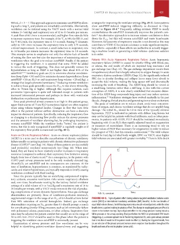
of 0.4 should achieve better FIGURE 49-5. Signs of patient effort during volume-targeted ventilation (volume assist-
supplemental oxygen in the range of an Fi O 2
than 90% saturation of arterial hemoglobin. Indeed, gas exchange control [VACV] or intermittent mandatory ventilation [IMV] breaths). In the two breaths of
greater than 0.5 should prompt a search
abnormalities requiring an Fi O 2 equal tidal volume shown, the left tracing represents a muscle-relaxed patient, while the other
for complicating alveolar filling processes, such as left ventricular failure breath shows a patient making inspiratory effort. The change in esophageal pressure (Peso) is
with pulmonary edema, pneumonia, or lobar collapse. Inspiratory flow shown in the bottom tracings. Signs of patient effort in the airway pressure tracing include a
rates may be adjusted for patient comfort but usually are in the range of fall in pressure at the airway opening (Pao) just before the VACV or synchronized IMV breath
50 to 60 L/min. PEEP should be used in this phase when the patient is (triggering, a), a concave upward rise in Pao during inspiration (b), and a peak airway pressure
triggering the ventilator since autoPEEP is universally present. that is less than it would be if the patient made no effort (c). During the triggered breath, Peso
Examination of airway pressure and flow waveforms can be very (as an indicator of the pleural pressure) remains more negative than baseline throughout the
helpful in identifying patient-ventilator dyssynchrony and suggesting breath and even after end inspiration (arrow at d).
section04.indd 431 1/23/2015 2:19:21 PM