
Page 612 - Hall et al (2015) Principles of Critical Care-McGraw-Hill
P. 612
432 PART 4: Pulmonary Disorders
that lung protection can be achieved using pressure-targeted ventilation,
for example as practiced in the Lung Open Ventilation Trial. Whatever
54
the mode, the respiratory rate should be set at 24 to 36 breaths per
minute as long as there is no autoPEEP. An occasional consequence of
lung protective ventilation is hypercapnia. This approach of preferring
hypercapnia to alveolar overdistention (“permissive hypercapnia”) is .
discussed further in Chaps. 51 and 55. 55,56 V
The Patient With Restriction of the Lungs or Chest Wall: A number of
restrictive diseases of the lungs or chest wall can lead to respiratory
failure, especially when there is a superimposed ventilatory challenge
(eg, pneumonia). These conditions are fully discussed in Chaps. 58, 86,
and 114 and include lung disease (eg, advanced pulmonary fibrosis or Time
late-stage ARDS), abdominal disease (eg, massive ascites), and other
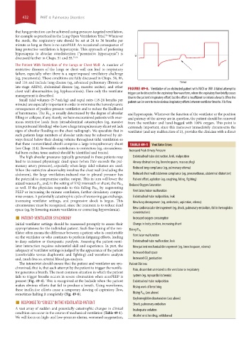
chest wall abnormalities (eg, kyphoscoliosis). Here only the ventilator FIGURE 49-6. Ventilation of an obstructed patient with VACV or IMV. A failed attempt to
trigger can be detected in the expiratory flow waveform, where the expiratory flow briefly ceases
management is described.
Small tidal volumes (5-7 mL/kg) and rapid rates (18-24 breaths per (due to the patient’s inspiratory effort) but the effort is insufficient to initiate a breath. Often the
minute) are especially important in order to minimize the hemodynamic patient can be seen to make obvious inspiratory efforts between ventilator breaths. V ˙ is flow.
consequences of positive-pressure ventilation and to reduce the likelihood
is usually determined by the degree of alveolar
of barotrauma. The Fi O 2 and hypercapnia. Whenever the function of the ventilator or the position
filling or collapse, if any. Rarely, we have encountered patients with enor- and patency of the airway are in question, the patient should be removed
mous restrictive loads from intraabdominal catastrophes (eg, massive from the ventilator and hand-bagged with 100% oxygen. This point is
intraperitoneal bleeding) who have a large intrapulmonary shunt yet lack extremely important, since this maneuver immediately circumvents the
signs of alveolar flooding on the chest radiograph. We speculate that in ventilator (and any malfunction of it), provides the clinician with a direct
such patients large numbers of alveolar units may be subserved by air-
ways forced below their closing volume throughout tidal ventilation so
that these nonventilated alveoli comprise a large intrapulmonary shunt TABLE 49-1 Ventilator Crises
(see Chap. 114). Reversible contributors to restriction (eg, circumferen-
tial burn eschar, tense ascites) should be identified and treated. Increased Peak Airway Pressure
The high alveolar pressures typically generated in these patients may Endotracheal tube obstruction, kink, malposition
lead to increased physiologic dead space (when Palv exceeds the pul- Airway obstruction (eg, bronchospasm, mucous plug)
monary artery pressure), especially when large tidal volumes are used.
When the restrictive abnormality involves the chest wall (including the Reduced lung compliance (eg, pulmonary edema)
abdomen), the large ventilation-induced rise in pleural pressure has Reduced chest wall/abdomen compliance (eg, pneumothorax, abdominal distention)
the potential to compromise cardiac output. This in turn will lower the Patient effort, agitation (eg, coughing, biting, fighting)
mixed venous P O 2 and, in the setting of V ˙ /Q ˙ mismatch or shunt, the Pa O 2 Reduced Oxygen Saturation
by augmenting
as well. If the physician responds to this falling Pa O 2
PEEP or increasing the minute ventilation, further circulatory compro- Ventilator/mixer malfunction
mise ensues. A potentially catastrophic cycle of worsening gas exchange, Endotracheal tube malposition, leak
increasing ventilator settings, and progressive shock is begun. This New lung derangement (eg, atelectasis, aspiration, edema)
circumstance must be recognized, since the treatment is to reduce dead
space (eg, by lowering minute ventilation or correcting hypovolemia). New cardiovascular derangement (eg, shock, pulmonary embolism, fall in hemoglobin
■ PATIENT-VENTILATOR SYNCHRONY Increased oxygen consumption
concentration)
Initial ventilator settings should be reassessed promptly to assess their Change in body position, increasing shunt
appropriateness for the individual patient. Such fine-tuning of the ven-
tilator often means the difference between a patient who is comfortable Rising P CO 2
on the ventilator or who continues to perform fatiguing efforts, leading Ventilator malfunction
to deep sedation or therapeutic paralysis. Assessing the patient-venti- Endotracheal tube malfunction, leak
lator interaction requires substantial skill and experience. In part, the New patient mechanical derangement (eg, bronchospasm, edema)
adequacy of ventilator settings is judged by the appearance of the patient
(comfortable versus diaphoretic and fighting) and waveform analysis Increased dead space
and, much less so, arterial blood gas analysis. Increased CO production
2
The intensivist should ensure that the patient and ventilator are syn- Patient Distress
chronized, that is, that each attempt by the patient to trigger the ventila- Pain, discomfort unrelated to the ventilator or respiratory
tor generates a breath. The most common situation in which the patient
fails to trigger breaths occurs in severe obstruction when autoPEEP is system (eg, myocardial ischemia)
present (Fig. 49-4). This is recognized at the bedside when the patient Endotracheal tube malposition
makes obvious efforts that fail to produce a breath. Using waveforms, Rising work of breathing
these ineffective efforts cause a temporary slowing of expiratory flow,
sometimes halting it completely (Fig. 49-6). Rising P CO 2 (see above)
■ RESPONSE TO “CRISES” IN THE VENTILATED PATIENT Oxyhemoglobin desaturation (see above)
A vast array of sudden and potentially catastrophic changes in clinical Shock, pulmonary embolism
Inadequate sedation
condition can occur in the course of mechanical ventilation (Table 49-1).
We will focus on high- and low-pressure alarms, worsened oxygenation, Alcohol or other drug, withdrawal
section04.indd 432 1/23/2015 2:19:22 PM