
Page 645 - Hall et al (2015) Principles of Critical Care-McGraw-Hill
P. 645
464 PART 4: Pulmonary Disorders
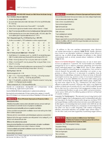
TABLE 52-9 NIH NHLBI ARDS Network Low-Tidal-Volume Ventilation Strategy TABLE 52-11 Contraindications to Permissive Hypercapnia and Respiratory Acidosis
Part I. Ventilator setup and adjustment Increased intracranial pressure from any cause (trauma, mass lesion, malignant hypertension)
1. Calculate ideal body weight (IBW). a Acute cerebrovascular disorders (eg, stroke)
2. Use assist/control mode and set initial tidal volume (VT) to 8 mL/kg IBW (if baseline Acute or chronic myocardial ischemia
VT >8 mL/kg). Severe pulmonary hypertension
3. Reduce VT by 1-mL/kg intervals every 2 hours until VT = 6 mL/kg IBW. Right ventricular failure
4. Set initial rate to approximate baseline minute ventilation (but not >35 bpm). Uncorrected severe metabolic acidosis
5. Adjust VT and respiratory rate (RR) to achieve pH and plateau pressure (Pplat) goals listed below. Sickle-cell anemia
6. Set the inspiratory flow rate above patient demand (usually >80 L/min); adjust flow Tricyclic antidepressant overdose
rate to achieve goal of inspiratory:expiratory ratio of 1:1.0-1.3
Patients taking β-blockers
= 88%-95%
Part II. Oxygenation goal: Pa O 2 =55-80 mm Hg or Sp O 2
Pregnancy (potential for decreased fetal blood flow due to vasodilatation-induced steal
-PEEP combinations to achieve oxygenation goal:
1. Use these incremental Fi O 2 syndrome; in addition, shift to the right of the oxygen dissociation curve may decrease
0.3 0.4 0.4 0.5 0.5 0.6 0.7 0.7 maternal-fetal oxygen gradient)
Fi O 2
PEEP 5 5 8 8 10 10 10 12
0.7 0.8 0.9 0.9 0.9 1.0 1.0 1.0
Fi O 2
In addition to this core ventilator management, some clinicians
PEEP 14 14 14 16 18 20 22 24
use various interventions as adjuncts (Table 52-12). Finally, clinicians
Part III. Plateau pressure (Pplat) Goal: = 30 cm H O may choose to use alternative ventilatory strategies whose efficacy is
2
, total RR, VT, and arterial blood gases unproven (eg, so-called salvage therapies for patients in dire clinical
1. Check Pplat (use 0.5-s inspiratory pause), Sp O 2
(ABG) (if available) at least every 4 hours and after each change in PEEP or VT. circumstances such as airway pressure-release ventilation or bilevel)
(Table 52-13).
2. If Pplat >30 cm H O, decrease VT by 1-mL/kg steps (minimum 4 mL/kg IBW).
2
3. If Pplat <25 cm H O and VT <6 mL/kg, increase VT by 1 mL/kg until Pplat >25 cm Adjuncts to Core Ventilator Management Clinicians may use one or more inter-
2
H O or VT = 6 mL/kg. ventions as adjuncts to “customize” the recommended core ventilator
2 management to try to improve pulmonary physiology and otherwise
4. If Pplat <30 cm H O and breath stacking occurs, one may increase VT in 1-mL/kg IBW benefit individual patients (see Table 52-13). Most of these adjuncts
2
increments (to a maximum of 8 mL/kg) as long as Pplat <30 cm H O.
2 hold the possibility of benefit based on extrapolation from animal or
Part IV. pH Goal: 7.30-7.45 clinical research that generally uses physiologic end points as sug-
Acidosis management: pH <7.30 gestions of efficacy. However, it is inaccurate to extrapolate from an
improvement in physiologic outcomes to efficacy in terms of clinically
<25 mm Hg (maximum
1. If pH = 7.15 −7.30, increase RR until pH >7.30 or Pa CO 2 meaningful outcomes (eg, survival or days of mechanical ventilation).
<25 mm Hg, may give NaHCO .
3 For example, in the first ARDSNet RCT, the lower-tidal-volume group
RR = 35); if RR = 35 and Pa CO 2
2. If pH <7.15 and NaHCO considered or infused, VT may be increased in 1-mL/kg steps had better survival than the higher-tidal-volume group, despite hav-
3
until pH >7.15 (Pplat goal may be exceeded). . Furthermore, the safety of these
3
ing lower mean values for Pa O 2 : Fi O 2
Alkalosis management: pH >7.45: Decrease RR if possible. adjuncts is generally uncertain. Clinicians who want to use high-level
evidence to guide care of patients with ALI and ARDS should be warned
a Male IBW = 50 + 2.3 (height [inches] − 60); female IBW = 45.5 + 2.3 (height [inches] − 60). ABG,
, oxygen saturation by pulse oximetry. that to date, save for a conservative fluid-management strategy, the use
arterial blood gas; SpO 2
of neuromuscular blockade in early severe ARDS, and prone position-
From the NIH NHLBI ARDS Network (complete protocol is available at www.ardsnet.org).
ing for severe ARDS, all of the following adjuncts fall short of that level
Reproduced with permission from Lanken PN. Acute respiratory distress syndrome. In: Lanken PN, of scientific evidence.
Hanson CW III, Manaker S, eds. The Intensive Care Unit Manual. Philadelphia, PA: WB Saunders; 2001:828.
Conservative (“Dry”) Fluid Management The rationale for restricting fluids in ALI
and ARDS suggests that if edemagenesis could be diminished early after
TABLE 52-10 Effects of Permissive Hypercapnia and Respiratory Acidosis
the lung injury, the duration of potentially dangerous ventilator, PEEP,
Hemodynamic effects and oxygen therapy could be reduced and outcome conceivably improved.
Activation of sympathetic nervous system and catechol secretion, normally resulting in In this regard, it is interesting that most patients with ARDS do not die
increased heart rate and stroke volume with peripheral vasodilatation during the early phase of disease as a consequence of severe hypoxemia,
but rather over days to weeks, frequently with evidence of hyperme-
Impaired myocardial contractility and worse congestive heart failure
tabolism, nosocomial infection, and multiple organ system failure. 73,74
Coronary vasodilation and potential for vasodilation-induced steal resulting in less blood On the other hand, some have argued that maximizing oxygen delivery
flow to ischemic myocardium
Pulmonary arteriolar vasoconstriction, potentiating hypoxic pulmonary vasoconstriction
Rightward shift of the oxygen dissociation curve with potential for less loading of O at the TABLE 52-12 Adjuncts to Consider in Addition to Low-Tidal-Volume
2
alveolar level and arterial desaturation Ventilation for Treatment of Acute Lung Injury (ALI) and Acute
Effects on the central nervous system Respiratory Distress Syndrome (ARDS)
Increased cerebral blood flow due to arterial hypertension and cerebral vasodilatation Conservative (“dry”) fluid management strategy
(vasodilatation may be lost in areas subject to trauma or ischemia) Early physical therapy
Cerebral vasodilation and potential vasodilation-induced steal resulting in less blood flow Early neuromuscular blocking agents (cisatracurium besylate)
to ischemic regions of the brain
Permissive hypercapnia
Increases intracranial pressure
Prone positioning
Data from Feihl F, Perret C. Permissive hypercapnia: how permissive should we be? Am J Respir Crit Care Recruitment maneuvers
Med. December 1994;150(6 pt 1):1722-1737.
section04.indd 464 1/23/2015 2:19:47 PM