
Page 720 - Hall et al (2015) Principles of Critical Care-McGraw-Hill
P. 720
CHAPTER 60: Liberation From Mechanical Ventilation 539
An SBT is deemed to fail if the patient develops tachypnea, hypox- Thus cessation of PPV or PEEP may lead to atelectasis and hypoxemia
emia, tachycardia, hypertension, new encephalopathy or arrhythmia, (Table 60-2). Hypoxemia can also result from cardiovascular changes
or signs of overt respiratory distress. In this case, ventilation should during weaning, so we are particularly vigilant to treat hypervolemia.
be resumed and, after attending to causes of failure, tried again in 24 Cardiovascular bases for failure: The transition to unassisted breathing is
hours. Those who sustain spontaneous breathing without failure can be associated with increased preload and left ventricular afterload which, com-
assessed for extubation. bined with hypervolemia, catecholamine secretion, and coronary artery
49
No study has examined the discriminative characteristics of ele- disease, may predispose to cardiogenic pulmonary edema. 50-55 Fluids often
ments used to judge success or failure of the SBT. Moreover, SBT modes administered during initial resuscitation—that can well exceed 5 to 10 L in
themselves may promote false- positive and false-negative results. Some some patients 29,56 —are redistributed from the third space and peripheral
patients who fail daily pressure support trials may be successfully extu- vasculature to the central circulation (Fig. 60-3). In patients with normal
bated after a brief T-piece, and the converse is also true. While the SBT hearts, these circulatory changes are usually well tolerated. However, in
40
38
is the final test of patients’ readiness (ie, few clinicians would extubate patients with left ventricular dysfunction, augmentation of preload and
after favorable weaning parameters alone without requiring a passed increased left ventricular afterload raise left ventricular work substantially.
SBT), no test is perfect, and liberation remains as much art as science. In addition, increased cardiac loads during weaning may precipitate isch-
■ THE PATIENT WHO FAILS INITIAL SBTS emia in those with coronary artery disease. Thus, the transition from PPV
to spontaneous breathing is accompanied by multiple events which can
Failure of an SBT is a clinical diagnosis. The signs of failure include rapid- contribute to left ventricular failure and cardiogenic pulmonary edema (see
shallow breathing, tachycardia (>110/minute), hypertension (increment Fig. 60-3). In 93 medical patients being weaned from mechanical ventila-
of >20 mm Hg), mental status changes, and subjective distress. These tion, ST-segment changes were noted in 6% of all patients, and in 10% of
signs result from (1) gas exchange failure, (2) circulatory decompensa- those with a preceding history of coronary artery disease. Weaning-related
55
tion, or (3) other issues. Although such patients often appear anxious, ischemia tended to increase the risk of weaning failure. Continuous moni-
anxiety is rarely the proximate cause of SBT failure. Arterial blood gas toring of ST-segments and treatment with additional nitrates may also be
analysis should not be used routinely to judge success or failure of an SBT. helpful in patients who experience ischemia during weaning.
When a patient fails an SBT, full ventilation should be resumed, usu- Ventilator and circuit factors: The ventilator and its circuitry can
ally until the next day. Meanwhile, clinicians should focus on patient contribute to weaning failure by two mechanisms: (1) by increasing
factors, rather than ventilator settings, seeking treatable bases for failure respiratory loads during a spontaneous breathing trial enough to fatigue
as described below. For carefully selected patients with COPD who fail the respiratory muscles, and (2) by imposing significant, unrecognized
an SBT, noninvasive ventilation can be used as a bridge to extubation, respiratory muscle work during “rest” periods. The resistance of the
as described below. endotracheal tube increases with time, and this increase can occasionally
Pulmonary bases for failure: Many factors reduce respiratory muscle be of sufficient magnitude to impede weaning. Even during periods of
strength or increase respiratory muscle loads in critically ill patients. intended rest, the ventilator circuit can load 57,58 and covertly fatigue the
In patients who fail due to strength-load imbalance, we assess neuro- respiratory muscles, especially when insufficient flow or pressure is pro-
muscular function and the elements of respiratory load so as to identify vided. Patient-ventilator synchrony during rest periods reduces the like-
59
reversible elements. lihood that the ventilator is contributing to weaning failure. Irregular
Respiratory muscle weakness may reflect preexisting illness, but
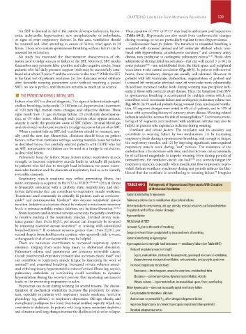
more commonly is acquired in the ICU as VIDD. 13,32,41,42 Critical illness TABLE 60-2 Pathogenesis of Hypoxemia and Hypercapnia With Cessation
is frequently associated with a catabolic state, malnutrition, and elec-
trolyte deficiencies that can contribute to respiratory muscle weakness. of Mechanical Ventilation
Treatments used commonly in critically ill patients such as corticoste- Factors Contributing to Hypoxemia
roids and neuromuscular blockers also depress respiratory muscle Pulmonary edema due to mobilization of peripheral edema
44
43
function. Sedatives and opiates should be reduced to minimum necessary Atelectasis due to recumbency, old age, obesity, residual sedatives, surfactant depletion
levels to enhance mobility, reduce delirium, and facilitate liberation. 18,19,21 (in patients with diffuse alveolar damage)
Bronchospasm and increased airways secretions frequently contribute
to resistive loading of the respiratory muscles. Elevated airway resis- Hypoventilation
tance greater than 15 cm H O/L per second can frequently be reversed Withdrawal of PEEP
2
by removing excessive airway secretion or treating with aerosolized Increased V ˙ due to the work of breathing
45
bronchodilators. If resistance remains greater than 15 cm H O/L per O 2
46
2
second despite bronchodilators in a patient who repeatedly fails to wean, Congestive heart failure precipitated by increased work of breathing
a therapeutic trial of corticosteroids may be helpful. Factors Contributing to Hypercapnia
There are numerous contributors to increased respiratory system Hypercapnia due to strength-load imbalance = ventilatory failure (see Table 60-3)
elastance, ranging from acute lung injury to abdominal distention.
Pulmonary edema and pneumonia are common reversible causes. Reduced respiratory muscle strength
Occult positive end-expiratory pressure also increases elastic load and Sepsis, malnutrition, electrolyte derangements, prolonged mechanical ventilation,
47
can contribute to respiratory muscle fatigue by increasing the work of dyssynchronous mechanical ventilation, corticosteroids, postparalytic syndrome
assisted and unassisted breathing. Increased minute volumes associ- Increased respiratory muscle loads
48
ated with lung injury, hypermetabolic states of critical illness (eg, sepsis),
pulmonary embolism, or overfeeding could contribute to dynamic Resistance—bronchospasm, excessive secretions, endotracheal tube
hyperinflation during the recovery process, thus increasing mechanical Elastance—pulmonary edema, dynamic hyperinflation, obesity
loads on the recovering respiratory muscles. Minute volume—hypermetabolism, increased dead space, fever, overfeeding
Hypoxemia can occur during weaning for several reasons. The discon-
tinuation of mechanical ventilation increases the propensity for atelec- Other hypercapnia—does not necessarily signal ventilatory failure
tasis, especially in patients with respiratory muscle weakness, restrictive Compensation for metabolic alkalosis
physiology (eg, obesity), or respiratory depression. Old age, obesity, and Acute return to premorbid P CO 2 after iatrogenic hyperventilation
recumbency predispose to a lower functional residual capacity, which can Hyperoxic hypercapnia (in chronic hypercapnic respiratory failure patients)
contribute to atelectasis. In patients with lung injury, surfactant depletion
and ultrastructural lung changes increase the likelihood of alveolar collapse. Residual sedatives/narcotics
section04.indd 539 1/23/2015 2:20:44 PM