
Page 848 - Hall et al (2015) Principles of Critical Care-McGraw-Hill
P. 848
CHAPTER 65: Pneumonia 579
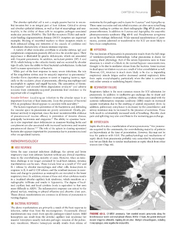
The alveolar epithelial cell is not a simply passive barrier to micro- exotoxins by the pathogen and is classic for S aureus and Aspergillus sp.
18
bial invasion but is an integral part of host defense. Critical to airway These same exotoxins and microbial enzymes can also cause actual lung
and alveolar epithelial defense, as well as that of macrophage and neu- necrosis, leading to cavities on chest radiographs (Fig. 65-2) and even
trophils, is the ability of these cells to recognize pathogen-associated pneumothoraxes. In addition to S aureus and Aspergillus, the anaerobic
molecular patterns (PAMPs). The Toll-like receptors (TLRs) and nucle- pleuropneumonia syndrome (Fig. 65-1) and Pseudomonas aeruginosa
otide-binding oligomerization domain (NOD) receptors are important are in the etiologic differential. While unusual manifestations of pneu-
examples. Binding of microbial markers to these receptors results in monia, patients admitted or transferred to the ICU are more likely to
both enhanced killing and initiation of the cascade of cytokines and have these complications.
chemokines characteristic of innate immune response.
A variety of other molecules contribute to alveolar defense. IgG and ■ HYPOXEMIA
complement components passively diffuse into the alveolar space. Their The mechanism of hypoxemia in pneumonia results from the full range
importance is illustrated by genetic deficiencies, which are associated of ventilation/perfusion relationships. Lobar pneumonia is classic for
with frequent pneumonia. In addition, surfactant protein (SP)-A and causing shunt physiology. Part of the severe hypoxemia seen in these
SP-D, which belong to the collectin family and are secreted by alveolar situations is a result of a block in the normal hypoxic vasoconstriction,
type-II cells, have the ability to bind bacterial carbohydrates. Surfactant thought to be due to mediator release from the bacteria. Some increase
itself increases opsonization of bacteria. in dead space ventilation occurs as a result of lobar consolidation as well.
While part of the host response in many severe infections, the role However, CO retention is much more likely to be due to impending
2
of the coagulation system may be uniquely important in pneumonia. respiratory muscle fatigue and/or decreased central respiratory drive
11
Alveolar fibrin deposition appears to assist in trapping bacteria, espe- from septic encephalopathy, particularly when the latter is combined
cially in the exudative phase of pneumonia, allowing macrophages and with either uremia or underlying hepatic disease.
neutrophils to capture and engulf bacteria. The association of throm-
bocytopenia and elevated fibrin degradation products and adverse ■ RESPIRATORY FAILURE
13
12
outcome from community-acquired pneumonia lend credence to the
important role of coagulation. Respiratory failure is the most common reason for ICU admission for
pneumonia. In addition to ineffective gas exchange due to shunt and
Vascular Defense: Localizing an infection to the alveolar space is an ventilation/perfusion mismatching, cytokine release associated with the
important function of host immunity. Even the presence of bacterial systemic inflammatory response syndrome (SIRS) results in increased
DNA in peripheral blood appears to correlate with mortality. 14 minute ventilation due to the resetting of central respiratory drive. In
The factors important in prevention of the bacteremia and defending addition, pulmonary compliance is decreased by the consolidation and
the vascular space in pneumonia are poorly understood. Clearly, pre- airway resistance may be increased by the presence of secretions. These
formed antibody is important, since the most incontrovertible evidence both result in substantially increased work of breathing. Pleuritic chest
of pneumococcal vaccine efficacy is prevention of invasive disease, pain and splinting may also contribute to the worsening gas exchange.
principally bacteremia and empyema. The ability to opsonize bacte- ■
15
ria is also important since deficiencies in mannose-binding lectin and HYPOTENSION
complement are also associated with increased bacteremia and invasive Septic shock is also a manifestation of severe pneumonia. For pneumo-
19
pneumococcal disease. 16,17 The role of the spleen in clearing opsonized nia acquired in the community, the overwhelming majority of patients
bacteria also appears important for pneumonia due to pneumococci and are hypovolemic at the time of presentation. However, this may not be
other encapsulated bacteria. true for patients with HAP. The pathophysiology and mechanisms of
septic shock from pneumonia probably vary somewhat by microorgan-
PATHOPHYSIOLOGY ism but are likely due to similar mechanisms as septic shock from other
■ HOST RESPONSE sources (see Chap. 64).
Given the near constant infectious challenge, the airway and lower
respiratory tract host defenses function without any clinical manifesta-
tions in the overwhelming majority of cases. However, when an infec-
tious challenge is no longer contained by local host defense, systemic
manifestations can be seen. These include fever as a result of TNF and
IL-1 release by alveolar macrophages and other cells, leukocytosis in
response to IL-6 and G-CSF release, and the resultant increase in secre-
tions and change to purulence as neutrophils are recruited to the lower
respiratory tract. In addition, release of these and other cytokines results
in a localized alveolar-capillary leak syndrome, which manifests as a
radiographic infiltrate and results in hypoxemia. The degree of local-
ized capillary leak and local cytokine levels is equivalent to that seen
more diffusely in ARDS. The inflammatory response can extend to the
pleural surface, resulting in pleural effusions and pleuritic chest pain.
These clinical manifestations are common in all forms of pneumonia to
varying degrees.
■ BACTERIAL RESPONSES
The above manifestations are primarily a result of the host response to
infection, rather than from the microorganism. Occasionally clinical
manifestations may result from specific pathogen-related factors. Mild FIGURE 65-2. CA-MRSA pneumonia. Note rounded necrotic pneumonia along the
hemoptysis can result from the alveolar capillary leak syndrome but bronchovascular bundle and small pleural effusion. Within 12 hours, the patient developed
massive hemoptysis usually indicates pathogen invasion of the pulmo- massive empyema ultimately requiring decortication. Multiple echocardiograms, including
nary vasculature. Massive hemoptysis usually results from release of transesophageal, were negative for endocarditis.
section05_c61-73.indd 579 1/23/2015 12:47:55 PM