
Page 186 - Clinical Anatomy
P. 186
ECA3 7/18/06 6:45 PM Page 171
The bones and joints of the upper limb 171
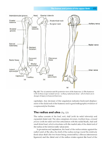
Fig. 122◊The (a) anterior and (b) posterior view of the humerus. (c) The humerus
with its three major related nerves—axillary, radial and ulnar—all of which are in
danger of injury in humeral fractures.
capitulum. Any decrease of this angulation indicates backward displace-
ment of the distal end of the humerus and is good radiographic evidence of
a supracondylar fracture.
The radius and ulna (Fig. 123)
The radius consists of the head, neck, shaft (with its radial tuberosity) and
expanded distal end. The ulna comprises olecranon, trochlear fossa, coronoid
process (with its radial notch for articulation with the radial head), shaft and
small distal head, which articulates with the medial side of the distal end of
the radius at the inferior radio-ulnar joint.
In pronation and supination, the head of the radius rotates against the
radial notch of the ulna, the shaft of the radius swings round the relatively
fixed ulnar shaft (the two bones being connected by a fibrous interosseous
ligament) and the distal end of the radius rotates against the head of the