
Page 185 - Clinical Anatomy
P. 185
ECA3 7/18/06 6:45 PM Page 170
170 The upper limb
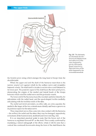
Fig. 121◊The deformity
of a fractured clavicle—
downward displacement
and adduction of the
outer fragment by
gravity and muscle
spasm respectively;
slight elevation of the
inner fragment by the
sternocleidomastoid.
the bicipital groove along which emerges the long head of biceps from the
shoulder joint.
Where the upper end and the shaft of the humerus meet there is the
narrow surgical neck against which lie the axillary nerve and circumflex
humeral vessels. The shaft itself is circular in section above and flattened in
its lower part. The posterior aspect of the shaft bears the faint spiral groove,
demarcating the origins of the medial and lateral heads of the triceps
between which wind the radial nerve and the profunda vessels.
The lower end of the humerus bears the rounded capitulum laterally, for
articulation with the radial head, and the spool-shaped trochlea medially,
articulating with the trochlear notch of the ulna.
The medial and lateral epicondyles, on either side, are extra-capsular; the
medial is the larger of the two, extends more distally and bears a groove on
its posterior aspect for the ulnar nerve.
Three important nerves thus come into close contact with the humerus
—the axillary, the radial and the ulnar; they may be damaged, respectively,
in fractures of the humeral neck, midshaft and lower end (Fig. 122).
It is an important practical point to note that the lower end of the
humerus is angulated forward 45° on the shaft. This is easily confirmed by
examining a lateral radiograph of the elbow, when it will be seen that a
vertical line continued downwards along the front of the shaft bisects the