
Page 191 - Clinical Anatomy
P. 191
ECA3 7/18/06 6:45 PM Page 176
176 The upper limb
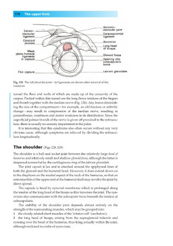
Fig. 128◊The left shoulder joint—its ligaments are shown after removal of the
humerus.
tunnel the floor and walls of which are made up of the concavity of the
carpus. Packed within this tunnel are the long flexor tendons of the fingers
and thumb together with the median nerve (Fig. 126). Any lesion diminish-
ing the size of the compartment— for example, an old fracture or arthritic
change —may result in compression of the median nerve, resulting in
paraesthesiae, numbness and motor weakness in its distribution. Since the
superficial palmar branch of the nerve is given off proximal to the retinacu-
lum, there is usually no sensory impairment in the palm.
It is interesting that this syndrome also often occurs without any very
obvious cause, although symptoms are relieved by dividing the retinacu-
lum longitudinally.
The shoulder (Figs 128, 129)
The shoulder is a ball-and-socket joint between the relatively large head of
humerus and relatively small and shallow glenoid fossa, although the latter is
deepened somewhat by the cartilaginous ring of the labrum glenoidale.
The joint capsule is lax and is attached around the epiphyseal lines of
both the glenoid and the humeral head. However, it does extend down on
to the diaphysis on the medial aspect of the neck of the humerus, so that an
osteomyelitis of the upper end of the humeral shaft may involve the joint by
direct spread.
The capsule is lined by synovial membrane which is prolonged along
the tendon of the long head of the biceps as this traverses the joint. The syn-
ovium also communicates with the subscapular bursa beneath the tendon of
subscapularis.
The stability of the shoulder joint depends almost entirely on the
strength of the surrounding muscles, which may be grouped into:
1◊◊the closely related short muscles of the ‘rotator cuff’ (see below);
2◊◊the long head of biceps, arising from the supraglenoid tubercle and
crossing over the head of the humerus, thus lying actually within the joint,
although enclosed in a tube of synovium;