
Page 189 - Clinical Anatomy
P. 189
ECA3 7/18/06 6:45 PM Page 174
174 The upper limb
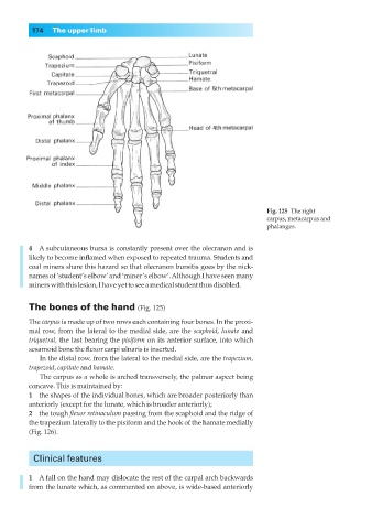
Fig. 125◊The right
carpus, metacarpus and
phalanges.
4◊◊A subcutaneous bursa is constantly present over the olecranon and is
likely to become inflamed when exposed to repeated trauma. Students and
coal miners share this hazard so that olecranon bursitis goes by the nick-
names of ‘student’s elbow’ and ‘miner’s elbow’. Although I have seen many
miners with this lesion, I have yet to see a medical student thus disabled.
The bones of the hand (Fig. 125)
The carpus is made up of two rows each containing four bones. In the proxi-
mal row, from the lateral to the medial side, are the scaphoid, lunate and
triquetral, the last bearing the pisiform on its anterior surface, into which
sesamoid bone the flexor carpi ulnaris is inserted.
In the distal row, from the lateral to the medial side, are the trapezium,
trapezoid, capitate and hamate.
The carpus as a whole is arched transversely, the palmar aspect being
concave. This is maintained by:
1◊◊the shapes of the individual bones, which are broader posteriorly than
anteriorly (except for the lunate, which is broader anteriorly);
2◊◊the tough flexor retinaculum passing from the scaphoid and the ridge of
the trapezium laterally to the pisiform and the hook of the hamate medially
(Fig. 126).
Clinical features
1◊◊A fall on the hand may dislocate the rest of the carpal arch backwards
from the lunate which, as commented on above, is wide-based anteriorly