
Page 187 - Clinical Anatomy
P. 187
ECA3 7/18/06 6:45 PM Page 172
172 The upper limb
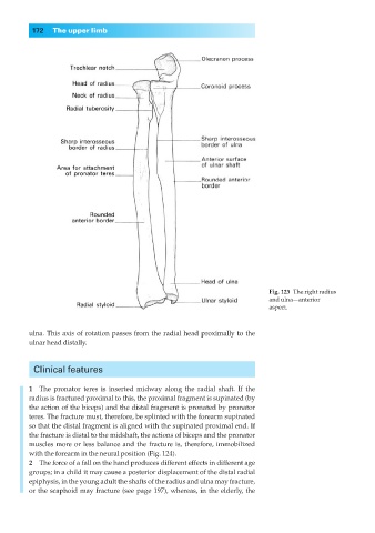
Fig. 123◊The right radius
and ulna—anterior
aspect.
ulna. This axis of rotation passes from the radial head proximally to the
ulnar head distally.
Clinical features
1◊◊The pronator teres is inserted midway along the radial shaft. If the
radius is fractured proximal to this, the proximal fragment is supinated (by
the action of the biceps) and the distal fragment is pronated by pronator
teres. The fracture must, therefore, be splinted with the forearm supinated
so that the distal fragment is aligned with the supinated proximal end. If
the fracture is distal to the midshaft, the actions of biceps and the pronator
muscles more or less balance and the fracture is, therefore, immobilized
with the forearm in the neural position (Fig. 124).
2◊◊The force of a fall on the hand produces different effects in different age
groups; in a child it may cause a posterior displacement of the distal radial
epiphysis, in the young adult the shafts of the radius and ulna may fracture,
or the scaphoid may fracture (see page 197), whereas, in the elderly, the