
Page 234 - Clinical Anatomy
P. 234
ECA4 7/18/06 6:47 PM Page 219
The bones and joints of the lower limb 219
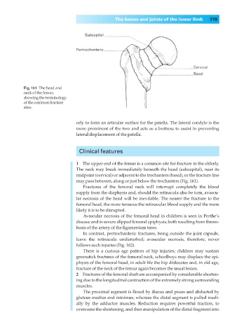
Fig. 161◊The head and
neck of the femur,
showing the terminology
of the common fracture
sites.
orly to form an articular surface for the patella. The lateral condyle is the
more prominent of the two and acts as a buttress to assist in preventing
lateral displacement of the patella.
Clinical features
1◊◊The upper end of the femur is a common site for fracture in the elderly.
The neck may break immediately beneath the head (subcapital), near its
midpoint (cervical) or adjacent to the trochanters (basal), or the fracture line
may pass between, along or just below the trochanters (Fig. 161).
Fractures of the femoral neck will interrupt completely the blood
supply from the diaphysis and, should the retinacula also be torn, avascu-
lar necrosis of the head will be inevitable. The nearer the fracture to the
femoral head, the more tenuous the retinacular blood supply and the more
likely it is to be disrupted.
Avascular necrosis of the femoral head in children is seen in Perthe’s
disease and in severe slipped femoral epiphysis; both resulting from throm-
bosis of the artery of the ligamentum teres.
In contrast, pertrochanteric fractures, being outside the joint capsule,
leave the retinacula undisturbed; avascular necrosis, therefore, never
follows such injuries (Fig. 162).
There is a curious age pattern of hip injuries; children may sustain
greenstick fractures of the femoral neck, schoolboys may displace the epi-
physis of the femoral head, in adult life the hip dislocates and, in old age,
fracture of the neck of the femur again becomes the usual lesion.
2◊◊Fractures of the femoral shaft are accompanied by considerable shorten-
ing due to the longitudinal contraction of the extremely strong surrounding
muscles.
The proximal segment is flexed by iliacus and psoas and abducted by
gluteus medius and minimus, whereas the distal segment is pulled medi-
ally by the adductor muscles. Reduction requires powerful traction, to
overcome the shortening, and then manipulation of the distal fragment into