
Page 235 - Clinical Anatomy
P. 235
ECA4 7/18/06 6:47 PM Page 220
220 The lower limb
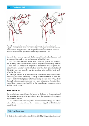
Fig. 162◊(a) Apertrochanteric fracture does not damage the retinacular blood
supply—aseptic bone necrosis does not occur. (b) Asubcapital fracture cuts off most
of the retinacular supply to the head—aseptic bone necrosis is common. Note that
the blood supply via the ligamentum teres is negligable in adult life.
line with the proximal segment; the limb must therefore be abducted and
also pushed forwards by using a large pad behind the knee.
Fractures of the lower end of the shaft, immediately above the condyles,
are relatively rare; fortunately so, because they may be extremely difficult
to treat since the small distal fragment is tilted backwards by gastrocne-
mius, the only muscle which is attached to it. The sharp proximal edge of
this distal fragment may also tear the popliteal artery, which lies directly
behind it (Fig. 163).
3◊◊The angle subtended by the femoral neck to the shaft may be decreased,
producing a coxa vara deformity. This may result from adduction fractures,
slipped the femoral epiphysis or bone-softening diseases. Coxa valga, where
the angle is increased, is much rarer but occurs in impacted abduction frac-
tures. Note, however, that in children the normal angle between the neck
and shaft is about 160°.
The patella
The patella is a sesamoid bone, the largest in the body, in the expansion of
the quadriceps tendon, which continues from the apex of the bone as the
ligamentum patellae.
The posterior surface of the patella is covered with cartilage and articu-
lates with the two femoral condyles by means of a larger lateral and smaller
medial facet.
Clinical features
1◊◊Lateral dislocation of the patella is resisted by the prominent articular