
Page 232 - Clinical Anatomy
P. 232
ECA4 7/18/06 6:47 PM Page 217
The bones and joints of the lower limb 217
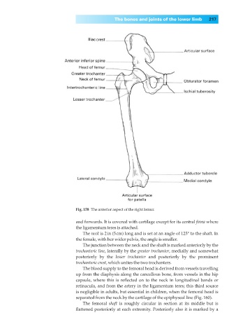
Fig. 158◊The anterior aspect of the right femur.
and forwards. It is covered with cartilage except for its central fovea where
the ligamentum teres is attached.
The neck is 2in (5cm) long and is set at an angle of 125° to the shaft. In
the female, with her wider pelvis, the angle is smaller.
The junction between the neck and the shaft is marked anteriorly by the
trochanteric line, laterally by the greater trochanter, medially and somewhat
posteriorly by the lesser trochanter and posteriorly by the prominent
trochanteric crest, which unites the two trochanters.
The blood supply to the femoral head is derived from vessels travelling
up from the diaphysis along the cancellous bone, from vessels in the hip
capsule, where this is reflected on to the neck in longitudinal bands or
retinacula, and from the artery in the ligamentum teres; this third source
is negligible in adults, but essential in children, when the femoral head is
separated from the neck by the cartilage of the epiphyseal line (Fig. 160).
The femoral shaft is roughly circular in section at its middle but is
flattened posteriorly at each extremity. Posteriorly also it is marked by a