
Page 407 - Clinical Anatomy
P. 407
ECA6 7/18/06 6:54 PM Page 392
392 The central nervous system
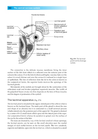
Fig. 274◊The lacrimal
gland and its drainage
system.
The conjunctiva is the delicate mucous membrane lining the inner
surface of the lids from which it is reflected over the anterior part of the
sclera to the cornea. Over the lids it is thick and highly vascular, but over the
sclera it is much thinner and over the cornea it is reduced to a single layer
of epithelium. The line of reflection from the lid to the sclera is known as
the conjunctival fornix; the superior fornix receives the openings of the
lacrimal glands.
Movements of the eyelids are brought about by the contraction of the
orbicularis oculi and levator palpebrae superioris muscles. The width of
the palpebral fissure at any one time depends on the tone of these muscles
and the degree of protrusion of the eyeball.
The lacrimal apparatus (Fig. 274)
The lacrimal gland is situated in the upper, lateral part of the orbit in what is
known as the lacrimal fossa. The main part of the gland is about the size
and shape of an almond, but it is connected to a small terminal process
which extends into the posterior part of the upper lid. The gland is drained
by a series of 8–12 small ducts which open into the lateral part of the supe-
rior conjunctival fornix whence its secretion is spread over the surface of
the eye by the action of the lids.
The tears are drained by way of the lacrimal canaliculi whose openings,
the lacrimal puncta, can be seen on the small elevation near the medial
margin of each eyelid known as the lacrimal papilla. The two canaliculi,
superior and inferior, open into the lacrimal sac, which is situated in a small