
Page 406 - Clinical Anatomy
P. 406
ECA6 7/18/06 6:54 PM Page 391
The special senses 391
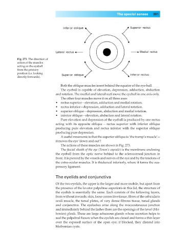
Fig. 273◊The direction of
action of the muscles
acting on the eyeball
from the primary
position (i.e. looking
directly forwards).
Both the oblique muscles insert behind the equator of the eye-ball.
The eyeball is capable of elevation, depression, adduction, abduction
and rotation. The medial and lateral recti move the eyeball in one axis only.
The other four muscles move it on all three axes:
•◊◊rectus superior—elevation, adduction and medial rotation.
•◊◊rectus inferior—depression, adduction and lateral rotation.
•◊◊superior oblique—depression, abduction and medial rotation.
•◊◊inferior oblique—elevation, abduction and lateral rotation.
Pure elevation and depression of the eyeball is produced by one rectus
acting with its opposite oblique — rectus superior with inferior oblique
producing pure elevation and rectus inferior with the superior oblique
producing pure depression.
Auseful mnemonic is that the superior oblique is ‘the tramp’s muscle’—
it moves the eye ‘down and out’!
The actions of these muscles are shown in Fig. 273.
The fascial sheath of the eye (Tenon’s capsule) is the membrane enclosing
the eyeball from the optic nerve behind to the sclerocorneal junction in
front. It is pierced by the vessels and nerves of the eye and by the tendons of
the extra-ocular muscles. It is thickened inferiorly, where it forms the sus-
pensory ligament.
The eyelids and conjunctiva
Of the two eyelids, the upper is the larger and more mobile, but apart from
the presence of the levator palpebrae superioris in this lid, the structure of
the eyelids is essentially the same. Each consists of the following layers,
from without inwards: skin, loose connective tissue, fibres of the orbicularis
oculi muscle, the tarsal plates, of very dense fibrous tissue, tarsal glands
and conjunctiva. The eyelashes arise along the mucocutaneous junction
and immediately behind the lashes there are the openings of the tarsal (Mei-
bomian) glands. These are large sebaceous glands whose secretion helps to
seal the palpebral fissure when the eyelids are closed and forms a thin layer
over the exposed surface of the open eye; if blocked, they distend into
Meibomian cysts.