
Page 60 - The Netter Collection of Medical Illustrations - Integumentary System_ Volume 4 ( PDFDrive )
P. 60
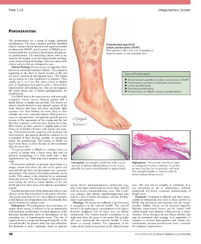
Plate 2-33                                                                                            Integumentary System


       POROKERATOSIS


       The  porokeratoses  are  a  group  of  benign  epidermal
       proliferations.  The  most  common  and  best-described           Disseminated superficial
       clinical variants include disseminated superficial actinic        actinic porokeratosis (DSAP).
       porokeratosis (DSAP), porokeratosis of Mibelli, poro-             Thin patches with a thin rim of epidermal
       keratosis palmaris et plantaris disseminata, and punctu-          hyperkeratosis on sun-exposed skin
       ate porokeratosis. The underlying disease state is the
       same for all variants, as are the characteristic and diag-
       nostic histopathological findings. There are many other
       clinical variants that are infrequently seen.
         Clinical Findings: Porokeratoses are typically inher-
       ited in an autosomal dominant fashion. They manifest
       beginning  in  the  third  to  fourth  decades  of  life  and                  Types of Porokeratosis
       are  more  common  in  sun-exposed  areas.  The  lesions
       can be minute to a few centimeters in diameter. They                             Disseminated superficial actinic porokeratosis (DSAP)
       usually  are  1-  to  2-cm,  thin,  flesh-colored  to  slightly                  Porokeratosis palmaris et plantaris disseminata
       pink  or  hyperpigmented  patches  with  a  characteristic                       Linear porokeratosis
       hyperkeratotic surrounding rim. This rim encompasses                             Punctate porokeratosis
       the  entire  lesion  and  is  almost  pathognomonic  for                         Porokeratosis of Mibelli (solitary porokeratosis)
       porokeratosis.
         The DSAP form is the most common and most easily
       recognized  clinical  variant.  Patients  present  with  a
       family history of similar skin growths. The lesions are
       almost  entirely  located  in  sun-exposed  regions  of  the
       body.  Patients  who  have  had  more  ultraviolet  light
       exposure  over  their  lifetime  are  more  likely  to  have
       multiple and more noticeable lesions. Most porokera-
       toses are asymptomatic, and patients typically present
       because of the appearance of the lesions and the fact
       that they continue to develop more lesions over time.
       Most lesions are flesh colored to slightly pink or red.
       Some can be frankly inflamed, with redness and crust-
       ing. Transformation into squamous cell carcinoma has
       been reported. and patients should be counseled to be
       reevaluated  if  they  develop  growths  or  ulcerations
       within  the  porokeratosis.  The  lesions  of  DSAP  are
       much more likely to affect the skin on the extremities
       than the facial skin.
         The porokeratosis of Mibelli is a solitary lesion, or
       a  group  of  lesions  with  a  linear  array  that  have  an
       identical  morphology  of  a  thin  patch  with  a  thin
       hyperkeratotic rim. They may occur anywhere on the
       body.                                      Low power. An atrophic epidermis with a scant  High power. The cornoid lamella is made
         Porokeratosis  palmaris  et  plantaris  disseminata  is  a   amount of dermal inflammation is seen. A peri-  of compacted stratum corneum. It overlies
       unique  variant  that  affects  the  skin  of  the  palms  and   pherally located cornoid lamella is appreciated.  an area that has lost its granular cell layer.
       soles initially and then can disseminate into a general-                            The cornoid lamella is oriented with its
       ized pattern. The lesions of the palms and soles can be                             distal ends pointing inward.
       tender. This variant is also inherited in an autosomal
       dominant manner. The lesions begin on the palms and
       soles  during  the  third  to  fourth  decades  of  life  and
       slowly spread to other areas of the skin in a generalized   taking  chronic  immunosuppressive  medications  (e.g.,   seen.  The  area  can  be  atrophic  or  acanthotic.  It  is
       pattern.                                  after solid organ transplantation) and in those infected   not  uncommon  to  see  an  inflammatory  infiltrate
         Punctate porokeratosis of the palms and soles is a rare   with the human immunodeficiency virus. This is indi-  underneath  the  lesion,  composed  predominantly  of
       clinical variant that is localized to the palms and soles.   rect  evidence  that  chronic  immunosuppression  may   lymphocytes.
       The lesions tend to be 0.5 to 1 cm in diameter and have   lead to a lack of tumor surveillance and the develop-  Treatment:  Treatment  is  difficult  and  often  unsuc-
       a well-defined rim of hyperkeratosis. Occasionally, they   ment of porokeratosis.   cessful for widespread areas such as those involved in
       can be mistaken for plantar warts.          Histology: On biopsy, the hallmark of porokeratosis   DSAP.  Sun  protection  and  sunscreen  use  are  recom-
         Pathogenesis: The pathogenesis of porokeratosis, no   is  recognition  of  the  cornoid  lamella.  The  cornoid   mended.  Solitary  lesions  can  be  removed  surgically.
       matter which variant, is believed to be an abnormality   lamella is the pathological representation of the hyper-  Multiple  disseminated  lesions  can  be  ablated  with
       of keratinocyte proliferation. A clonal expansion of the   keratotic  peripheral  rim  of  tissue  seen  on  clinical   carbon dioxide laser ablation, 5-fluorouracil, or derm-
       abnormal  keratinocytes  leads  to  development  of  the   examination.  The  cornoid  lamella  is  positioned  at  an   abrasion. These therapies are not always effective and
       expanding  rim  of  hyperkeratotic  tissue.  This  rim  of   angle away from the center of the lesion. The granular   may  be  associated  with  scarring.  It  is  imperative  to
       hyperkeratosis is recognized histopathologically as the   cell  layer  underneath  the  cornoid  lamella  is  often   continue  to  monitor  these  patients  with  routine  skin
       cornoid lamella. No genetic defect has been identified.   absent  or  severely  thinned.  The  appearance  of  the   examinations,  because  porokeratoses  have  a  potential
       Porokeratosis  is  more  commonly  found  in  patients   center of the lesion is dependent on the clinical variant   for malignant degeneration.

       46                                                                                    THE NETTER COLLECTION OF MEDICAL ILLUSTRATIONS