
Page 59 - The Netter Collection of Medical Illustrations - Integumentary System_ Volume 4 ( PDFDrive )
P. 59
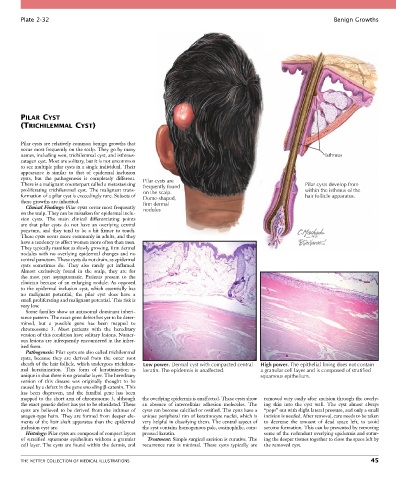
Plate 2-32                                                                                                  Benign Growths


        PILAR CYST
        (TRICHILEMMAL CYST)


        Pilar cysts are relatively common benign growths that
        occur most frequently on the scalp. They go by many
        names, including wen, trichilemmal cyst, and isthmus-                                                    Isthmus
        catagen cyst. Most are solitary, but it is not uncommon
        to see multiple pilar cysts in a single individual. Their
        appearance  is  similar  to  that  of  epidermal  inclusion
        cysts,  but  the  pathogenesis  is  completely  different.   Pilar cysts are
        There is a malignant counterpart called a metastasizing   frequently found                        Pilar cysts develop from
        proliferating  trichilemmal  cyst.  The  malignant  trans-  on the scalp.                         within the isthmus of the
        formation of a pilar cyst is exceedingly rare. Subsets of   Dome-shaped,                          hair follicle apparatus.
        these growths are inherited.              firm dermal
          Clinical Findings: Pilar cysts occur most frequently   nodules
        on the scalp. They can be mistaken for epidermal inclu-
        sion  cysts.  The  main  clinical  differentiating  points
        are  that  pilar  cysts  do  not  have  an  overlying  central
        punctum,  and  they  tend  to  be  a  bit  firmer  to  touch.
        These cysts occur more commonly in adults, and they
        have a tendency to affect women more often than men.
        They typically manifest as slowly growing, firm dermal
        nodules  with  no  overlying  epidermal  changes  and  no
        central punctum. These cysts do not drain, as epidermal
        cysts  sometimes  do.  They  also  rarely  get  inflamed.
        Almost  exclusively  found  in  the  scalp,  they  are  for
        the  most  part  asymptomatic.  Patients  present  to  the
        clinician  because  of  an  enlarging  nodule.  As  opposed
        to  the  epidermal  inclusion  cyst,  which  essentially  has
        no  malignant  potential,  the  pilar  cyst  does  have  a
        small proliferating and malignant potential. This risk is
        very low.
          Some families show an autosomal dominant inheri-
        tance pattern. The exact gene defect has yet to be deter-
        mined,  but  a  possible  gene  has  been  mapped  to
        chromosome  3.  Most  patients  with  the  hereditary
        version of this condition have solitary lesions. Numer-
        ous lesions are infrequently encountered in the inher-
        ited form.
          Pathogenesis: Pilar cysts are also called trichilemmal
        cysts,  because  they  are  derived  from  the  outer  root
        sheath of the hair follicle, which undergoes trichilem-  Low power. Dermal cyst with compacted central  High power. The epithelial lining does not contain
        mal  keratinization.  This  form  of  keratinization  is   keratin. The epidermis is unaffected.  a granular cell layer and is composed of stratified
        unique in that there is no granular layer. The hereditary                          squamous epithelium.
        version  of  this  disease  was  originally  thought  to  be
        caused by a defect in the gene encoding β-catenin. This
        has  been  disproven,  and  the  familial  gene  has  been
        mapped to the short arm of chromosome 3, although   the overlying epidermis is unaffected. These cysts show   removed very easily after excision through the overly-
        the exact genetic defect has yet to be elucidated. These   an  absence  of  intercellular  adhesion  molecules.  The   ing  skin  into  the  cyst  wall.  The  cyst  almost  always
        cysts  are  believed  to  be  derived  from  the  isthmus  of   cysts can become calcified or ossified. The cysts have a   “pops” out with slight lateral pressure, and only a small
        anagen-type hairs. They are formed from deeper ele-  unique peripheral rim of keratinocyte nuclei, which is   incision is needed. After removal, care needs to be taken
        ments  of  the  hair  shaft  apparatus  than  the  epidermal   very helpful in classifying them. The central aspect of   to  decrease  the  amount  of  dead  space  left,  to  avoid
        inclusion cyst are.                       the cyst contains homogenous pale, eosinophilic, com-  seroma formation. This can be prevented by removing
          Histology: Pilar cysts are composed of compact layers   pressed keratin.          some of the redundant overlying epidermis and sutur-
        of  stratified  squamous  epithelium  without  a  granular   Treatment: Simple surgical excision is curative. The   ing the deeper tissues together to close the space left by
        cell layer. The cysts are found within the dermis, and   recurrence  rate  is  minimal.  These  cysts  typically  are   the removed cyst.


        THE NETTER COLLECTION OF MEDICAL ILLUSTRATIONS                                                                           45