
Page 61 - The Netter Collection of Medical Illustrations - Integumentary System_ Volume 4 ( PDFDrive )
P. 61
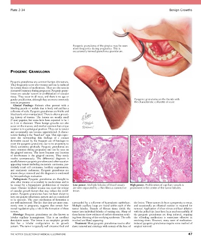
Plate 2-34                                                                                                  Benign Growths


                                                   Pyogenic granuloma of the gingiva may be seen
                                                   more frequently during pregnancy. This is
                                                   occasionally termed granuloma gravidarum.


        PYOGENIC GRANULOMA


        Pyogenic granulomas are common benign skin tumors.
        They frequently occur after trauma and can be induced
        by certain classes of medications. They are also seen in
        increased frequency during pregnancy. Pyogenic granu-
        lomas are vascular tumors or proliferations of vascular
        tissue. They occur in all races, and there is no age or
        gender predilection, although they are more commonly                                Pyogenic granuloma on the thumb with
        seen in pregnancy.                                                                  the characteristic collarette of scale
          Clinical  Findings:  Patients  often  present  with  a
        bleeding papule or nodule that is beefy red and has a
        collarette of scale. Pyogenic granulomas are friable and
        bleed easily when manipulated. There is often a preced-
        ing  history  of  trauma.  The  lesions  are  usually  small
        (5 mm) papules, but some have been reported to be 1
        to  2 cm  in  diameter.  These  benign  growths  can  also
        occur on the mucosa, and another common but unique
        location is in a periungual position. They can be tender
        and occasionally can become superinfected. A charac-
        teristic finding is the “band-aid” sign. This sign repre-
        sents  the  surrounding  skin  findings  of  a  contact
        dermatitis  caused  by  the  frequent  use  of  bandages  to
        cover the pyogenic granuloma due to its propensity to
        bleed,  sometimes  profusely.  Pyogenic  granulomas  are
        more common during pregnancy and can be seen on
        the gingival mucosa. The most frequent oral location
        of  involvement  is  the  gingival  mucosa.  They  rarely
        resolve  spontaneously.  The  differential  diagnosis  is
        usually between pyogenic granuloma and other vascular-
        appearing tumors including metastatic carcinoma, par-
        ticularly  renal  cell  carcinoma,  bacillary  angiomatosis,
        and  amelanotic  melanoma.  Pyogenic  granulomas  are
        almost always removed and the diagnosis is confirmed
        by histopathologic evaluation.
          Pathogenesis:  Pyogenic  granulomas  are  thought  to
        arise after trauma or secondary to medications and to
        be  caused  by  a  hyperplastic  proliferation  of  vascular   Low power. Multiple lobules of blood vessels   High power. Proliferation of capillary vessels is
        tissue. Chronic localized trauma can cause the release   are seen separated by a thin fibrous connective  prominent in the center of the tumor lobules.
        of vascular growth factors that may induce the prolif-  tissue.
        eration. Pyogenic granulomas have not been shown to
        have any genetic inheritance pattern and are considered
        to be sporadic. The exact mechanism of formation is
        not well understood. The fact that they are more com-  surrounded by a collarette of hyperplastic epithelium.   the lesion. These tumors do have a propensity to recur,
        monly  seen  in  pregnancy  suggests  that  certain  hor-  Multiple capillary loops are found within each of the   and  occasionally  an  elliptical  excision  is  required  for
        monal regulations play a role in the formation of these   tumor  lobules.  Strands  of  fibrous  tissue  divide  the   removal. Application of silver nitrate and laser ablation
        tumors.                                   tumor into individual lobules of varying size. Many of   with the pulsed dye laser have been used successfully. If
          Histology:  Pyogenic  granulomas  are  also  known  as   these lesions show evidence of surface ulceration result-  the  pyogenic  granulomas  are  drug  induced,  stopping
        lobular  capillary  hemangiomas.  This  is  an  excellent   ing from thinning of the overlying epidermis. The cells   the  offending  medication  is  sometimes  effective  in
        descriptive  name.  The  lesion  is  an  exophytic  growth   involved are bland appearing.  resolving  them.  However,  many  cases  of  medication-
        that  has  a  lobular  configuration  to  its  growth     Treatment: Most pyogenic granulomas resolve after   induced pyogenic granulomas require some method of
        pattern. The tumor is typically well circumscribed and   shave removal and curettage with cautery of the base of   surgical removal.


        THE NETTER COLLECTION OF MEDICAL ILLUSTRATIONS                                                                           47